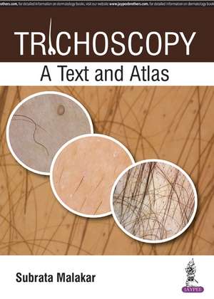
Trichoscopy: A Text and Atlas
Autor Subrata Malakaren Limba Engleză Hardback – 16 iul 2017
This book is a step by step guide to trichoscopy for practising dermatologists. Beginning with an overview of devices and tools, and trichoscopic terminologies, the following sections cover the diagnostic imaging of many different hair and scalp disorders, including alopecia, hair weathering, infection and infestation, psoriasis, and more. Complete sections are dedicated to systemic diseases and paediatric hair disorders.
The book concludes with algorithms to help diagnose different disorders, and discussion on monitoring and follow up.
The practical text is further enhanced with nearly 600 images to assist learning and self assessment.
Key points
- Step by step guide to trichoscopic imaging for diagnosis of hair and scalp disorders
- Covers numerous disorders and includes section on paediatric trichoscopy
- Features algorithms to assist diagnosis
- Highly illustrated with nearly 600 clinical images
Preț: 1439.10 lei
Preț vechi: 1514.85 lei
-5% Nou
275.36€ • 288.28$ • 227.85£
Carte tipărită la comandă
Livrare economică 02-08 aprilie
Specificații
ISBN-10: 9386150743
Pagini: 400
Dimensiuni: 216 x 279 mm
Greutate: 1.6 kg
Editura: JAYPEE BROTHERS MEDICAL PUBLISHERS PVT LTD
Colecția Jaypee Brothers,Medical Publishers Pvt. Ltd.
Locul publicării:Delhi, India
Cuprins
Section 1: Devices and Tools
- Introduction
Section 2: Devices and Tools
- Devices and Tools
Section 3: Trichoscopic Terminologies
- Trichoscopic Terminologies
Section 4: Trichoscopic Language and Pattern Analysis
- Follicular and Perifollicular Patterns
- Interfollicular Pattern
- Pattern of Hair Shafts
- Pattern of Scaly Scalp
Section 5: Normal Scalp
- Normal Scalp
Section 6: Nonscarring Alopecias
- Androgenetic Alopecia
- Female Pattern Hair Loss
- Alopecia Areata Incognito
- Alopecia Areata
- Trichotillomania
- Traction Alopecia
- Telogen Effluvium
- Anagen Effluvium
Section 7: Scarring Alopecias
- Lichen Planopilaris
- Frontal Fibrosing Alopecia
- Discoid Lupus Erythematosus
- Acne Keloidalis Nuchae
- Central Centrifugal Cicatricial Alopecia
- Folliculitis Decalvans
- Dissecting Cellulitis
- Pseudopelade of Brocq
Section 8: Hair Weathering
- Trichoscopy of Damaged Hair
Section 9: Infection and Infestation
- Tinea Capitis
- Pediculosis Capitis
Section 10: Inflammatory Scalp Diseases
- Scalp Psoriasis, Seborrheic Dermatitis and Sebopsoriasis
Section 11: Systemic Diseases
- Trichoscopy in Autoimmune Bullous Disorders
- Trichoscopy in Autoimmune Connective Tissue Diseases
Section 12: Pediatric Trichology
- Pediatric Hair Disorders
Section 13: Body Hair Disorders
- Body Hair
Section 14: Effect of Dermatology Practice on Hair
- Trichoscopy in Hair Transplantation
Section 15: Algorithms
- Algorithm
Section 16: Monitoring Therapeutic Efficacy
- Trichoscopic Monitoring and Follow-up
Notă biografică
Subrata Malakar MBBS MD DCH
Director and Chief Dermatologist, Rita Skin Foundation, Kolkata, West Bengal, India
Descriere
Trichoscopy is the dermoscopic imaging of the scalp and hair. The method is based on dermoscopy and videodermoscopy and is used for the evaluation and diagnosis of hair and scalp diseases.
This book is a step by step guide to trichoscopy for practising dermatologists. Beginning with an overview of devices and tools, and trichoscopic terminologies, the following sections cover the diagnostic imaging of many different hair and scalp disorders, including alopecia, hair weathering, infection and infestation, psoriasis, and more. Complete sections are dedicated to systemic diseases and paediatric hair disorders.
The book concludes with algorithms to help diagnose different disorders, and discussion on monitoring and follow up.
The practical text is further enhanced with nearly 600 images to assist learning and self assessment.
Key points
- Step by step guide to trichoscopic imaging for diagnosis of hair and scalp disorders
- Covers numerous disorders and includes section on paediatric trichoscopy
- Features algorithms to assist diagnosis
- Highly illustrated with nearly 600 clinical images
