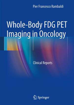
Whole-Body FDG PET Imaging in Oncology: Clinical Reports
Autor Pier Francesco Rambaldien Limba Engleză Paperback – 27 noi 2013
Preț: 382.06 lei
Preț vechi: 402.17 lei
-5% Nou
Puncte Express: 573
Preț estimativ în valută:
73.12€ • 76.05$ • 60.36£
73.12€ • 76.05$ • 60.36£
Carte disponibilă
Livrare economică 24 martie-07 aprilie
Preluare comenzi: 021 569.72.76
Specificații
ISBN-13: 9788847052949
ISBN-10: 8847052947
Pagini: 200
Ilustrații: XIX, 352 p. 256 illus., 229 illus. in color.
Dimensiuni: 178 x 254 x 17 mm
Greutate: 0.75 kg
Ediția:2014
Editura: Springer
Colecția Springer
Locul publicării:Milano, Italy
ISBN-10: 8847052947
Pagini: 200
Ilustrații: XIX, 352 p. 256 illus., 229 illus. in color.
Dimensiuni: 178 x 254 x 17 mm
Greutate: 0.75 kg
Ediția:2014
Editura: Springer
Colecția Springer
Locul publicării:Milano, Italy
Public țintă
Professional/practitionerCuprins
Galbladder and Biliary Ducts.- Head and Neck.- Colon and Rectum.- Oesophagus.- Gynecology.- Lymphomas and Thymomas.- Breast.- Melanoma.- Pancreas.- Lung.- Stomach.- Urinary Tract.
Recenzii
“This book provides to students and experts in nuclear medicine, radiology and oncology, but also to all other clinicians interested in better understanding the clinical role of PET/FDG in oncology, a guidance for the interpretation of the images, which correlate anatomical and functional data. … a clinically valuable report correctly answering the diagnostic query is obtained. … useful for all physicians who are in charge of oncological patients to contextualize, explain and communicate the results obtained with PET/CT … .” (Vania Mallardo, European Journal of Nuclear Medicine and Molecular Imaging, Vol. 42, 2015)
Textul de pe ultima copertă
This manual presents a large collection of clinical cases in oncology with accompanying whole-body FDG PET-CT scans. The aim is to promote an integrated approach to the use of PET-CT, and detailed attention is therefore paid to the clinical history and diagnostic question. A central aspect of every clinical case described in this manual is the guidance on the clinical report, which is the official tool for communicating with both the referring physician and the person undergoing the diagnostic test; for this reason it needs to be clear, understandable, and written in shared language. The advice regarding report preparation is strongly supported by informative PET, CT, and PET-CT fused images of each disease. The book is broadly structured according to anatomic region, and a wide range of common diseases likely to be imaged using PET-CT is covered. This book will be of value to all those training or working in the field of oncology who wish to ensure that they are best placed to contextualize, interpret, and report the findings obtained with PET-CT, which can have such a dramatic impact on prognosis, therapeutic choice, and quality of life.
Caracteristici
Large collection of clinical cases in oncology with accompanying whole-body FDG PET-CT scans Promotes an integrated approach to the use of PET-CT, with detailed attention to clinical history and the diagnostic question Offers extensive guidance on clinical report preparation Covers a wide range of diseases and disease locations?
