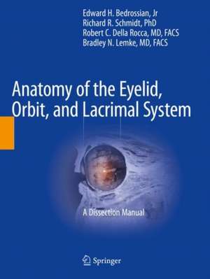
Anatomy of the Eyelid, Orbit, and Lacrimal System: A Dissection Manual
Editat de Edward H. Bedrossian, Jr, Richard R. Schmidt, Robert C. Della Rocca, Bradley N. Lemkeen Limba Engleză Paperback – 2 feb 2023
Opening chapters provide an introduction to the topic and outline instruments needed for the dissections. Subsequent chapters then describe the dissection of the eyelid in various layered approaches. Then, further discussions demonstrate the neuroanatomy of the cranial fossae, the cavernous sinus, and the dissection of deep orbital structures from an anterior, superior and lateral approach. Closing chapters then examine the nasolacrimal system and nasal cavities
Anatomy of the Eyelid, Orbit, and Lacrimal System is an expertly written invaluable resource for the surgeon seeking to enhance their knowledge and surgical skills.
| Toate formatele și edițiile | Preț | Express |
|---|---|---|
| Paperback (1) | 834.82 lei 6-8 săpt. | |
| Springer International Publishing – 2 feb 2023 | 834.82 lei 6-8 săpt. | |
| Hardback (1) | 1160.63 lei 3-5 săpt. | |
| Springer International Publishing – feb 2022 | 1160.63 lei 3-5 săpt. |
Preț: 834.82 lei
Preț vechi: 878.77 lei
-5% Nou
Puncte Express: 1252
Preț estimativ în valută:
159.74€ • 167.21$ • 132.96£
159.74€ • 167.21$ • 132.96£
Carte tipărită la comandă
Livrare economică 31 martie-14 aprilie
Preluare comenzi: 021 569.72.76
Specificații
ISBN-13: 9783030882679
ISBN-10: 3030882675
Pagini: 71
Ilustrații: XIII, 71 p. 231 illus., 230 illus. in color.
Dimensiuni: 210 x 279 mm
Greutate: 0.24 kg
Ediția:1st ed. 2022
Editura: Springer International Publishing
Colecția Springer
Locul publicării:Cham, Switzerland
ISBN-10: 3030882675
Pagini: 71
Ilustrații: XIII, 71 p. 231 illus., 230 illus. in color.
Dimensiuni: 210 x 279 mm
Greutate: 0.24 kg
Ediția:1st ed. 2022
Editura: Springer International Publishing
Colecția Springer
Locul publicării:Cham, Switzerland
Cuprins
1 . Preparation of Specimens for Orbital Dissection Course.- 2. The Eyelids.- 3. Anterior Orbit.- 4. Neuroanatomy: Cavernous Sinus.- 5. The Orbit: Superior Approach.- 6. The Orbit: Lateral Approach.- 7. Paranasal Sinuses and the Nasolacrimal Drainage System.
Notă biografică
Edward H. Bedrossian Jr., MD, FACS
Wills Eye Hospital
Sydney Kimmel Medical College of Thomas Jefferson University
Philadelphia , PA
Lewis Katz School of Medicine at Temple University
Philadelphia, PA
Wills Eye Hospital
Sydney Kimmel Medical College of Thomas Jefferson University
Philadelphia , PA
Lewis Katz School of Medicine at Temple University
Philadelphia, PA
Richard R. Schmidt, PhD
Sydney Kimmel Medical College of Thomas Jefferson University
Philadelphia, PA
USA
Associate Editors :
Robert C Della Rocca M.D. F.A.C.S.
Surgeon Director and Chief, Ophthalmic Plastic, Reconstructive and Orbital Surgery
New York Eye and Ear Infirmary of Mount Sinai
Clinical Professor of Ophthalmology,
Ichahn School of Medicine of Mount Sinai
New York. New York
Bradley N. Lemke,M.D. F.A.C.S.
Clinical Professor of Ophthalmology
Universityof Wisconsin School of Medicine and Public Health
Madison , Wisconsin
Textul de pe ultima copertă
This book is a dissection manual and atlas on the anatomy of the eyelid, orbit, and lacrimal system; it functions as a succinct yet comprehensive resource.
Opening chapters provide an introduction to the topic and outline instruments needed for the dissections. Subsequent chapters then describe the dissection of the eyelid in various layered approaches. Then, further discussions demonstrate the neuroanatomy of the cranial fossae, the cavernous sinus, and the dissection of deep orbital structures from an anterior, superior and lateral approach. Closing chapters then examine the nasolacrimal system and nasal cavities
Anatomy of the Eyelid, Orbit, and Lacrimal System is an expertly written invaluable resource for the surgeon seeking to enhance their knowledge and surgical skills.
Anatomy of the Eyelid, Orbit, and Lacrimal System is an expertly written invaluable resource for the surgeon seeking to enhance their knowledge and surgical skills.
Caracteristici
Features hundreds of high-quality color photos and line drawings Includes clinical correlation to highlight the importance of an individual anatomic structure Written by experts in the field
