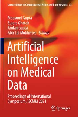
Artificial Intelligence on Medical Data: Proceedings of International Symposium, ISCMM 2021: Lecture Notes in Computational Vision and Biomechanics, cartea 37
Editat de Mousumi Gupta, Sujata Ghatak, Amlan Gupta, Abir Lal Mukherjeeen Limba Engleză Paperback – 24 iul 2023
| Toate formatele și edițiile | Preț | Express |
|---|---|---|
| Paperback (1) | 1168.30 lei 6-8 săpt. | |
| Springer Nature Singapore – 24 iul 2023 | 1168.30 lei 6-8 săpt. | |
| Hardback (1) | 1175.63 lei 6-8 săpt. | |
| Springer Nature Singapore – 24 iul 2022 | 1175.63 lei 6-8 săpt. |
Din seria Lecture Notes in Computational Vision and Biomechanics
- 15%
 Preț: 651.19 lei
 - 5%
 Preț: 734.38 lei
 - 5%
 Preț: 1103.03 lei
 - 5%
 Preț: 724.50 lei
 - 5%
 Preț: 714.63 lei
 - 20%
 Preț: 344.42 lei
 - 5%
 Preț: 720.10 lei
 - 5%
 Preț: 367.64 lei
 - 5%
 Preț: 731.43 lei
 - 15%
 Preț: 648.05 lei
 - 5%
 Preț: 725.07 lei
 - 5%
 Preț: 374.20 lei
 - 5%
 Preț: 735.11 lei
 - 5%
 Preț: 729.42 lei
 - 15%
 Preț: 647.40 lei
 - 5%
 Preț: 1100.30 lei
 - 5%
 Preț: 374.57 lei
 - 15%
 Preț: 642.68 lei
 - 5%
 Preț: 380.80 lei
 - 5%
 Preț: 375.70 lei
 - 24%
 Preț: 888.75 lei
 -
 Preț: 483.05 lei
 - 20%
 Preț: 569.86 lei
 - 15%
 Preț: 652.31 lei
 - 5%
 Preț: 647.55 lei
 -
 Preț: 355.15 lei
 - 18%
 Preț: 971.32 lei
 -
 Preț: 363.67 lei
 - 5%
 Preț: 1122.42 lei
 - 5%
 Preț: 1031.90 lei
 - 5%
 Preț: 1104.32 lei
 - 5%
 Preț: 721.77 lei
Preț: 1168.30 lei
Preț vechi: 1229.79 lei
-5% Nou
Puncte Express: 1752
Preț estimativ în valută:
223.58€ • 232.56$ • 184.58£
223.58€ • 232.56$ • 184.58£
Carte tipărită la comandă
Livrare economică 15-29 aprilie
Preluare comenzi: 021 569.72.76
Specificații
ISBN-13: 9789811901539
ISBN-10: 9811901538
Pagini: 484
Ilustrații: XII, 484 p. 262 illus., 194 illus. in color.
Dimensiuni: 155 x 235 mm
Greutate: 0.69 kg
Ediția:1st ed. 2023
Editura: Springer Nature Singapore
Colecția Springer
Seria Lecture Notes in Computational Vision and Biomechanics
Locul publicării:Singapore, Singapore
Cuprins
Retinal Vessel Segmentation in Fundus Image using Low-Cost Multiple U-Net Architecture.- Imaging Radiomic-Driven Framework for Automated Cancer Investigation.- Computational image analysis of painful and pain free intervertebral disc.- Bio-medical image encryption using the modified chaotic image encryption method.- Stability of Feature Selection Algorithms.- A Database Application of Monitoring Covid-19 in India.- Walking Assistant for Vision Impaired by Using Deep Learning.- Detection of Coronavirus in Electron Microscope Imagery using Convolutional Neural Networks.- IoT & RFID based Smart Card System Integrated with Healthcare, Electricity, QR and Banking Sectors.- Face Mask Recognition Using Modular, Wavelet and Modular-Wavelet PCA.- Effective Overview of Different ML Models used for Prediction of COVID -19 Patients.- Hybrid CNN-SVM Model for Face Mask Detector to Protect from COVID -19.- A Study on Various Medical Imaging Modalities and image fusion methods.- A Deep Learning Technique For Bi-fold Grading of an Eye Disorder DR-Diabetic Retinopathy.- Brain Tumor Detection Using Fine Tuned YOLO Model with Transfer Learning.
Notă biografică
Dr. Mousumi Gupta is an Associate professor of Computer Application at SMIT with more than a decade of teaching and research experience in the field of pattern recognition. She graduated with majoring in mathematics and did her masters in computer application. She further specialized in Information Technology with an M.Tech from Sikkim Manipal Institute of Technology (SMIT) in 2007. She completed her PhD from Sikkim Manipal University in 2015 during which she worked in the area of pattern recognition using digital image processing. She started her professional career after joining as a lecturer in the Department of Computer Science & Engineering in 2007 at SMIT. Her present research area include development of pattern recognition algorithm for digital pathology images. Presently, she has two research grants from ICMR and 26 peer reviewed publications.
Dr. Sujata Ghatak is an Assistant Professor of Computer Applications at Institute of Engineering & Management, India. She graduated from Calcutta University majoring in Computer Science following which she received Masters in Computer Science. She completed Master of Technology in Computer science from Sikkim Manipal Institute of Technology (SMIT), Majitar, Sikkim. She is one of the editor for Springer LNNS conference proceedings for IEMIS 2018. Her research interests include Image processing, Pattern Recognition, Applied Dynamical System in image processing, Natural Language Processing and Data Science. She is the member of several technical functional bodies such as The Society of Digital Information and Wireless Communications (SDIWC), Internet Society as a Global Member (ISOC), International Computer Science and Engineering Society (ICSES). She has chaired several sessions in various conferences in India. She has published several papers in reputed journals and conferences.
Dr. Amlan Gupta graduated in medical sciences from NMCH Patna in 1989 and Post-Graduated in Pathology from Calcutta University in 1995. After a brief interlude of private practice, he started his academic career in MCOMS Pokhara as an Assistant professor in 1997. He moved from there to SMIMS Gangtok and joined as an Associate professor in March of 2002. He became a professor in 2007. As a faculty he has been teaching, researching and taking care hospital diagnostics including surgical pathology and blood banking. His present area of research is wound healing and neural tissue healing. During his tenure he has 32 publications with 4 extramural research grants including one on quantitative histopathology.
Dr. Abir Lal Mukherjee is a board certified anatomic and clinical pathologist, working at Henry Ford Hospital, Detroit, Michigan as Chief of Clinical Neuropathology. He is a medical graduate from the oldest medical college in India, Calcutta Medical College. He post-graduated in pathology from JIPMER Pondicherry. He had further specialization in pathology at MD Anderson Cancer Center Houston Texas. He began his academic career in MCOMS Pokhara as assistant professor in 1996. He rose to the position of Associate Director, Pathology Residency Program & Director of Autopsy and Neuropathology, Department of Pathology and Laboratory Medicine, Temple University, School of Medicine, Philadelphia, PA. He is member of major scientific and educational committees, e.g., Graduate Medical Education Committee. He has 19 high impact publications. His primary research interests are in the area of Morphological spectrum of central nervous system malignancy and post mortem appearances of CNS in encephalitis and general incidence of undiagnosed malignancies in autopsy.
Textul de pe ultima copertă
This book includes high-quality papers presented at the Second International Symposium on Computer Vision and Machine Intelligence in Medical Image Analysis (ISCMM 2021), organized by Computer Applications Department, SMIT in collaboration with Department of Pathology, SMIMS, Sikkim, India, and funded by Indian Council of Medical Research, during 11 – 12 November 2021. It discusses common research problems and challenges in medical image analysis, such as deep learning methods. It also discusses how these theories can be applied to a broad range of application areas, including lung and chest x-ray, breast CAD, microscopy and pathology. The studies included mainly focus on the detection of events from biomedical signals.
Caracteristici
Covers latest trends in computer vision and hybrid machine learning paradigms for medical image analysis Presents magnetic resonance, computed tomography, optical and confocal microscopy, and video and range data images Serves as a reference for technologists, scientists, industry professionals, and researchers
