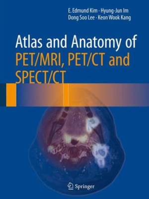
Atlas and Anatomy of PET/MRI, PET/CT and SPECT/CT
Autor E. Edmund Kim, Hyung-jun Im, Dong Soo Lee, Keon Wook Kangen Limba Engleză Hardback – 17 aug 2016
| Toate formatele și edițiile | Preț | Express |
|---|---|---|
| Paperback (1) | 1320.99 lei 6-8 săpt. | |
| Springer International Publishing – 12 iun 2018 | 1320.99 lei 6-8 săpt. | |
| Hardback (1) | 1695.39 lei 38-44 zile | |
| Springer – 17 aug 2016 | 1695.39 lei 38-44 zile |
Preț: 1695.39 lei
Preț vechi: 1784.62 lei
-5% Nou
Puncte Express: 2543
Preț estimativ în valută:
324.42€ • 339.41$ • 269.49£
324.42€ • 339.41$ • 269.49£
Carte tipărită la comandă
Livrare economică 29 martie-04 aprilie
Preluare comenzi: 021 569.72.76
Specificații
ISBN-13: 9783319286501
ISBN-10: 3319286501
Pagini: 189
Ilustrații: XI, 594 p. 582 illus. in color.
Dimensiuni: 210 x 279 x 38 mm
Greutate: 2.12 kg
Ediția:1st ed. 2016
Editura: Springer
Colecția Springer
Locul publicării:Cham, Switzerland
ISBN-10: 3319286501
Pagini: 189
Ilustrații: XI, 594 p. 582 illus. in color.
Dimensiuni: 210 x 279 x 38 mm
Greutate: 2.12 kg
Ediția:1st ed. 2016
Editura: Springer
Colecția Springer
Locul publicării:Cham, Switzerland
Public țintă
Professional/practitionerCuprins
Atlas and Anatomy of PET/MR .- Atlas and Anatomy of PET/CT.- Atlas and Anatomy of SPECT/CT.
Recenzii
“The book should be especially helpful to nuclear medicine and radiology residents, who may use it as a great source of ready-to-use knowledge in everyday clinical practice. It may also be useful to both undergraduate and postgraduate students, who will appreciate its importance as a good resource to improve their skill in interpreting hybrid nuclear medicine images. … very valuable in all imaging departments with hybrid machines in helping to improve the interpretation of individual cases in routine practice.” (Giuseppe Danilo Di Stasio and Luigi Mansi, European Journal of Nuclear Medicine and Molecular Imaging, Vol. 45, 2018)
Notă biografică
Edmund Kim, MD, MS Professor of Radiologic Sciences University of California at Irvine Irvine, CA 92697 Professor of Molecular Medicine Seoul National University and Kyunghee University Seoul, Korea Hyungjoon Im, MD, PhD Instructor Nuclear Medicine Department Seoul National University Hospital 101 Dae-Hak Ro Jong-Ro Gu Seoul, Korea 110-744 Dong-Soo Lee, MD, PhD Professor of Nuclear Medicine Nuclear Medicine Department Seoul National University Hospital 101 Dae-Hak Ro Jong-Ro Gu Seoul, Korea 110-744 Keon-Wook Kang, MD Professor and Chairman Nuclear Medicine Department Seoul National University Hospital 101 Dae-Hak Ro Jong-Ro Gu Seoul, Korea 110-744
Textul de pe ultima copertă
This atlas showcases cross-sectional anatomy for the proper interpretation of images generated from PET/MRI, PET/CT, and SPECT/CT applications. Hybrid imaging is at the forefront of nuclear and molecular imaging and enhances data acquisition for the purposes of diagnosis and treatment. Simultaneous evaluation of anatomic and metabolic information about normal and abnormal processes addresses complex clinical questions and raises the level of confidence of the scan interpretation. Extensively illustrated with high-resolution PET/MRI, PET/CT and SPECT/CT images, this atlas provides precise morphologic information for the whole body as well as for specific regions such as the head and neck, abdomen, and musculoskeletal system. Atlas and Anatomy of PET/MRI, PET/CT, AND SPECT/CT is a unique resource for physicians and residents in nuclear medicine, radiology, oncology, neurology, and cardiology.
Caracteristici
Integrates cross-sectional anatomy with PET/MRI, PET/CT, and SPECT/CT
Provides morphologic information via hybrid imaging for the whole body as well as for specific regions
Offers dedicated survey of anatomic and metabolic processes as visualized by PET/MRI
Includes supplementary material: sn.pub/extras
Provides morphologic information via hybrid imaging for the whole body as well as for specific regions
Offers dedicated survey of anatomic and metabolic processes as visualized by PET/MRI
Includes supplementary material: sn.pub/extras
