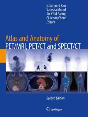
Atlas and Anatomy of PET/MRI, PET/CT and SPECT/CT
Editat de E. Edmund Kim, Vanessa Murad, Jin-Chul Paeng, Gi-Jeong Cheonen Limba Engleză Paperback – 5 feb 2023
| Toate formatele și edițiile | Preț | Express |
|---|---|---|
| Paperback (1) | 1293.75 lei 6-8 săpt. | |
| Springer International Publishing – 5 feb 2023 | 1293.75 lei 6-8 săpt. | |
| Hardback (1) | 1491.55 lei 38-44 zile | |
| Springer International Publishing – 5 feb 2022 | 1491.55 lei 38-44 zile |
Preț: 1293.75 lei
Preț vechi: 1361.84 lei
-5% Nou
Puncte Express: 1941
Preț estimativ în valută:
247.59€ • 268.84$ • 207.97£
247.59€ • 268.84$ • 207.97£
Carte tipărită la comandă
Livrare economică 22 aprilie-06 mai
Preluare comenzi: 021 569.72.76
Specificații
ISBN-13: 9783030923518
ISBN-10: 3030923517
Pagini: 281
Ilustrații: XI, 281 p. 266 illus., 264 illus. in color.
Dimensiuni: 210 x 279 mm
Greutate: 0.67 kg
Ediția:2nd ed. 2022
Editura: Springer International Publishing
Colecția Springer
Locul publicării:Cham, Switzerland
ISBN-10: 3030923517
Pagini: 281
Ilustrații: XI, 281 p. 266 illus., 264 illus. in color.
Dimensiuni: 210 x 279 mm
Greutate: 0.67 kg
Ediția:2nd ed. 2022
Editura: Springer International Publishing
Colecția Springer
Locul publicării:Cham, Switzerland
Cuprins
1. Atlas and Anatomy of PET/MRI.- 2. Atlas and Anatomy of PET/CT.- 3. Atlas and Anatomy of SPECT/CT.
Recenzii
“It is a book worth buying if you are at the beginning of your training in nuclear medicine and/or radiology, especially because of the analysis of the different scan slides in the same clinical case.” (Clara Ferreira, RAD Magazine, June, 2023)
Notă biografică
Dr. E. Edmund Kim was tenured Professor of Radiology and Medicine at the University of Texas M. D. Anderson Cancer Center for 31 years (1981-2012). He is also Professor of Radiological Sciences at the University of California at Irvine, as well as Professor of Molecular Medicine at Seoul National University in Korea. He has written more than 350 scientific papers and 15 books related to nuclear oncology and molecular imaging. He has been editor of Current Medical Imaging since 2005, and he was associate editor of the Journal of Nuclear Medicine and Molecular Imaging for 30 years.
Dr. Vanessa Murad is a Radiologist from the Department of Diagnostic Imaging at Fundacion Santa Fe de Bogota University Hospital in Colombia, as well as a clinical fellow from the Ko Chang-Soon Program at the Department of Nuclear Medicine at Seoul National University Hospital in Korea. Her main focus is oncological, hybrid and molecular imaging.
Dr. Jin Chul Paeng is a Nuclear Medicine physician and professor in the Department of Nuclear Medicine at Seoul National University Hospital (SNUH). He received his MD and PhD degrees from Seoul National University College of Medicine (SNUCM), and has worked mainly in the field of nuclear oncologic and nuclear cardiology, with many published papers. As a board-certified NM physician, he has worked in the Armed Forces Capital Hospital of Korea as Director of the Department of Nuclear Medicine. In 2008, he moved to SNUH to become a professor in charge of NM imaging, treatment, and radiation safety. His research fields include clinical application of NM imaging (particularly, in cardiac molecular imaging), and radiopharmaceutical treatment.
Dr. Gi-Jeong Cheon is a Nuclear Medicine physician, professor, and current chairman of the Department of Nuclear Medicine at the Seoul National University Hospital (SNUH) in Korea. He has worked mainly in the field of nuclear oncology and has published several original research papers. He manages more than 150 employees in Physics and Engineering, Chemistry, and Biology sections. The department operates PET/MRI, PET/CT, SPECT/CT, and SPECT with a new CZT crystal producing high-quality resolution.
Textul de pe ultima copertă
The Atlas and Anatomy of PET/MRI, PET/CT, AND SPECT/CT, Second Edition features cross-sectional anatomy to assist in proper interpretation of images generated from PET/MRI, PET/CT, and SPECT/CT applications. At the forefront of nuclear and molecular imaging, hybrid imaging improves data acquisition for diagnosis and treatment. Simultaneous evaluation of anatomic and metabolic information about normal and abnormal processes addresses complex clinical questions and elevates confidence in scan interpretations. Extensively illustrated with high-resolution PET/MRI, PET/CT and SPECT/CT images, the atlas offers precise morphologic information for the whole body, including specific regions such as the head and neck, abdomen, and musculoskeletal system. Updated with teaching cases and many recent non-FDG PET cases, Atlas and Anatomy of PET/MRI, PET/CT, AND SPECT/CT, Second Edition is a unique resource for physicians and residents in nuclear medicine, radiology, oncology, neurology, and cardiology.
Caracteristici
Integrates cross-sectional anatomy with PET/MRI, PET/CT, and SPECT/CT Provides morphologic information via hybrid imaging for the whole body as well as for specific regions Provides clinical correlation for each case with detailed imaging description
