Atlas of Clinical Cases on Brain Tumor Imaging
Editat de Yelda Özsunar, Utku Şenolen Limba Engleză Hardback – 29 apr 2020
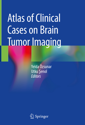
This book presents and analyzes clinical cases of brain tumors and follows the classification provided by the WHO in 2016. After introductory chapters reviewing the international literature on the topic, the advances made in all imaging modalities (especially Magnetic Resonance and Computed Tomography) are examined.
All radiological findings are supplemented with a wealth of images and brief explanations. The clinical information is given as part of the case discussion, as are the characteristics and differential diagnosis of the tumors. Radiologic-pathologic correlations round out the description of each clinical case.
Intended as a quick and illustrative reference guide for radiology residents and medical students, this atlas represents the most up-to-date, practice-oriented reference book in the field of Brain Tumor Imaging.
| Toate formatele și edițiile | Preț | Express |
|---|---|---|
| Paperback (1) | 611.64 lei 38-45 zile | |
| Springer International Publishing – 26 aug 2021 | 611.64 lei 38-45 zile | |
| Hardback (1) | 859.45 lei 38-45 zile | |
| Springer International Publishing – 29 apr 2020 | 859.45 lei 38-45 zile |
Preț: 859.45 lei
Preț vechi: 904.68 lei
-5% Nou
Puncte Express: 1289
Preț estimativ în valută:
164.45€ • 171.70$ • 136.11£
164.45€ • 171.70$ • 136.11£
Carte tipărită la comandă
Livrare economică 31 martie-07 aprilie
Preluare comenzi: 021 569.72.76
Specificații
ISBN-13: 9783030232726
ISBN-10: 3030232727
Pagini: 248
Ilustrații: VIII, 331 p. 238 illus., 80 illus. in color.
Dimensiuni: 178 x 254 mm
Greutate: 0.77 kg
Ediția:1st ed. 2020
Editura: Springer International Publishing
Colecția Springer
Locul publicării:Cham, Switzerland
ISBN-10: 3030232727
Pagini: 248
Ilustrații: VIII, 331 p. 238 illus., 80 illus. in color.
Dimensiuni: 178 x 254 mm
Greutate: 0.77 kg
Ediția:1st ed. 2020
Editura: Springer International Publishing
Colecția Springer
Locul publicării:Cham, Switzerland
Cuprins
PART I GENERAL CONSIDERATIONS IN BRAIN TUMORS.- Chapter 1 Pathology, Epidemiology and WHO Classification of Brain Tumors.- Chapter 2 When and How to Use Imaging in Brain Tumors, Protocols.- Chapter 3 Pearls in Conventional Imaging Methods for Brain Tumors.- Chapter 4 Diffusion, Perfusion and PET Imaging of Brain Tumors.- Chapter 5 Role of Magnetic Resonance Spectroscopy in Clinical Management of Brain Tumors.- Chapter 6 Diffusion Tensor Imaging and Functional Magnetic Resonance in Brain Tumor Imaging.- Chapter 7 Brain Tumours Imaging: Developing New Technique and Future Perspectives (Including Molecular Imaging, Radiogenomics).- Chapter 8 The Basic Molecular Genetics and the Common Mutations of Brain Tumors.- Chapter 9 Clinical Treatment of Brain Tumors.- PART II ~ CASE BASED LEARNING IN BRAIN TUMORS.- Chapter 10 Non neoplastic Mass Lesions of the Brain.- Chapter 11 Extraaxial Tumors.- Chapter 12 Pediatric BrainTumours.- Chapter 13 Primary Intra axial Brain Tumours.- Chapter 14 Secondary Brain Tumours.- Chapter 15 Tumour-like Conditions of the Brain .- Chapter 16 Preoperative Surgery Planning and Peroperative Imaging.- Chapter 17 Post-operative Imaging.- Chapter 18 Follow up and Treatment Changes.
Recenzii
“It appears clear that this book is more than only an Atlas of brain tumors. Being able to provide all the most important and characterizing neuroradiological data achievable in brain neoplasm using MRI (or CT), the publication has to be integrated with further information achievable with PET and hybrid imaging.” (Luigi Mansi, European Journal of Nuclear Medicine and Molecular Imaging, Vol. 48, 2021)
Notă biografică
Yelda Özsunar has been a Professor of Radiology at Adnan Menderes University School of Medicine since 2008. She graduated from Gazi University School of Medicine, Ankara, in 1992 and completed her radiology residency at the same university in 1998. During her radiology education, she studied at the University of Copenhagen, and after taking her specialty, she worked at Yale University’s Functional MR Imaging unit, New Haven and at Massachusettes General Hospital Neuroradiology Department Harvard Medical School, USA, as research fellow between 1999-2001. She is a European Board Certified Neuroradiologist and published over 75 international peer-reviewed articles and book chapters and has over 1500 international citations. She authored a popular science book called ‘Universal Symphony’ about electromagnetic spectrum and neuroscience.
Utku Şenol is Professor of Radiology at the Akdeniz University, School of Medicine, Antalya, Turkey, with full time faculty Chair at the Neuroradiology Department. He graduated in 1988 at the Faculty of Medicine at the Hacettepe University. After the residency in Radiology (1993) at the Akdeniz University, School of Medicine, he took his doctorate degree in Medical Informatics in 2013. He is member of several Turkish and International Societies, among them the European Society of Neuroradiology, the European Society of Radiology and the Radiological Society of North America, as well as Current President of the Turkish Medical Informatics Association and the TSR Imaging Informatics Working Group. His areas of interest are: Neuroradiology, MRI, Pediatric Neuroradiology, Medical Informatics, Imaging Informatics, Radiology Education, Radiology Management, Decision Support System, Evidence Based Medicine, and Radiology Physics.
Utku Şenol is Professor of Radiology at the Akdeniz University, School of Medicine, Antalya, Turkey, with full time faculty Chair at the Neuroradiology Department. He graduated in 1988 at the Faculty of Medicine at the Hacettepe University. After the residency in Radiology (1993) at the Akdeniz University, School of Medicine, he took his doctorate degree in Medical Informatics in 2013. He is member of several Turkish and International Societies, among them the European Society of Neuroradiology, the European Society of Radiology and the Radiological Society of North America, as well as Current President of the Turkish Medical Informatics Association and the TSR Imaging Informatics Working Group. His areas of interest are: Neuroradiology, MRI, Pediatric Neuroradiology, Medical Informatics, Imaging Informatics, Radiology Education, Radiology Management, Decision Support System, Evidence Based Medicine, and Radiology Physics.
Textul de pe ultima copertă
This book presents and analyzes clinical cases of brain tumors and follows the classification provided by the WHO in 2016. After introductory chapters reviewing the international literature on the topic, the advances made in all imaging modalities (especially Magnetic Resonance and Computed Tomography) are examined.
All radiological findings are supplemented with a wealth of images and brief explanations. The clinical information is given as part of the case discussion, as are the characteristics and differential diagnosis of the tumors. Radiologic-pathologic correlations round out the description of each clinical case.
Intended as a quick and illustrative reference guide for radiology residents and medical students, this atlas represents the most up-to-date, practice-oriented reference book in the field of Brain Tumor Imaging.
Caracteristici
Provides many imaging samples, together with brief and illustrative explanations Discusses numerous clinical tumor cases with an emphasis on advanced radiological imaging methods Contains principals and primary radiological findings on brain tumors
