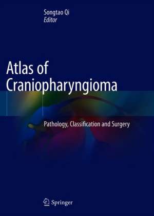
Atlas of Craniopharyngioma: Pathology, Classification and Surgery
Editat de Songtao Qien Limba Engleză Hardback – 28 noi 2019
This book covers histoembryology of craniopharyngioma, together with anatomical morphology and abundant clinical data, systematically showing an innovative classification method, i.e. QST classification. This classification method can better reflect the different origin and growth pattern of craniopharyngioma,the relationship between tumors and surrounding structure of the tumor growth pattern, and clinical significance in surgery. The 70 clinical cases with different classification and treatment history are discussed as an important reference for surgical treatment of craniopharyngioma. The anatomy, morphologyand pathology of sellar region also have great reference value for researchers in the field of neural science. The underlying intention of this book is to help bring a change in the concept that “craniopharyngioma is an incurable benign tumor only due to its anatomical location”.
| Toate formatele și edițiile | Preț | Express |
|---|---|---|
| Paperback (1) | 647.73 lei 38-44 zile | |
| Springer Nature Singapore – 29 noi 2020 | 647.73 lei 38-44 zile | |
| Hardback (1) | 905.07 lei 38-44 zile | |
| Springer Nature Singapore – 28 noi 2019 | 905.07 lei 38-44 zile |
Preț: 905.07 lei
Preț vechi: 952.71 lei
-5% Nou
Puncte Express: 1358
Preț estimativ în valută:
173.21€ • 178.69$ • 146.59£
173.21€ • 178.69$ • 146.59£
Carte tipărită la comandă
Livrare economică 28 februarie-06 martie
Preluare comenzi: 021 569.72.76
Specificații
ISBN-13: 9789811373213
ISBN-10: 9811373213
Pagini: 150
Ilustrații: XI, 167 p. 313 illus., 230 illus. in color.
Dimensiuni: 210 x 279 mm
Ediția:1st ed. 2020
Editura: Springer Nature Singapore
Colecția Springer
Locul publicării:Singapore, Singapore
ISBN-10: 9811373213
Pagini: 150
Ilustrații: XI, 167 p. 313 illus., 230 illus. in color.
Dimensiuni: 210 x 279 mm
Ediția:1st ed. 2020
Editura: Springer Nature Singapore
Colecția Springer
Locul publicării:Singapore, Singapore
Cuprins
1. Histology and embryology related to craniopharyngiomas.- 2. Surgical Anatomy.- 3. QST classification for craniopharyngioma and hispathological aspect.- 4. Comparison between the QST scheme and other schemes for craniopharyngiomas.- 5. Endoscopic transsphenoidal surgery for craniopharyngioma.- 6. Surgical treatment of craniopharyngioma: transcranial approach.- 7. Treatment of recurrent craniopharyngioma.- 8. Retreatment of craniopharyngioma after external radiotherapy and intracapsular radiochemotherapy.- 9. Basic research in craniopharyngioma.
Notă biografică
Songtao Qi is a professor of Neurosurgery Department of Nanfang Hospital, Southern Medical University, China. He is also the director at the Neurosurgery Department of Nanfang Hospital, Southern Medical University, China. Prof. Qi’s research focus is the effects of intracranial membrane structures, including dura, arachnoid and pia mater, on the development of intracranial diseases.
Textul de pe ultima copertă
This book aims to facilitate readers to understand the origin, growth pattern and relationship between tumor and adherent structure of craniopharyngioma, so as to improve the cure rate and safety of surgery. It’s contributed by Neurosurgery Department of Nanfang Hospital, Southern Medical University, China, which focuses on the management of craniopharyngioma.
This book covers histoembryology of craniopharyngioma, together with anatomical morphology and abundant clinical data, systematically showing an innovative classification method, i.e. QST classification. This classification method can better reflect the different origin and growth pattern of craniopharyngioma,the relationship between tumors and surrounding structure of the tumor growth pattern, and clinical significance in surgery. The 70 clinical cases with different classification and treatment history are discussed as an important reference for surgical treatment of craniopharyngioma. The anatomy, morphologyand pathology of sellar region also have great reference value for researchers in the field of neural science. The underlying intention of this book is to help bring a change in the concept that “craniopharyngioma is an incurable benign tumor only due to its anatomical location”.
This book covers histoembryology of craniopharyngioma, together with anatomical morphology and abundant clinical data, systematically showing an innovative classification method, i.e. QST classification. This classification method can better reflect the different origin and growth pattern of craniopharyngioma,the relationship between tumors and surrounding structure of the tumor growth pattern, and clinical significance in surgery. The 70 clinical cases with different classification and treatment history are discussed as an important reference for surgical treatment of craniopharyngioma. The anatomy, morphologyand pathology of sellar region also have great reference value for researchers in the field of neural science. The underlying intention of this book is to help bring a change in the concept that “craniopharyngioma is an incurable benign tumor only due to its anatomical location”.
Caracteristici
Facilitates readers to understand the complex disease of craniopharyngioma Discusses 70 clinical cases of craniopharyngioma Illustrates abundant pictures
