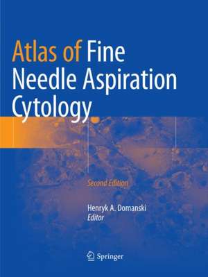
Atlas of Fine Needle Aspiration Cytology
Editat de Henryk A. Domanskien Limba Engleză Paperback – 22 dec 2018
With contributions from experts in the field internationally and many new colour images Atlas of Fine Needle Aspiration Cytology, Second Edition provides a comprehensive and up-to-date guide to FNAC for pathologists, cytopathologists, radiologists, oncologists, surgeons and others involved in the diagnosis and treatment of patients with suspicious mass lesions.
| Toate formatele și edițiile | Preț | Express |
|---|---|---|
| Paperback (2) | 1328.86 lei 38-44 zile | |
| SPRINGER LONDON – 29 sep 2016 | 1328.86 lei 38-44 zile | |
| Springer International Publishing – 22 dec 2018 | 1654.84 lei 38-44 zile | |
| Hardback (2) | 1654.37 lei 3-5 săpt. | |
| SPRINGER LONDON – 3 noi 2013 | 1654.37 lei 3-5 săpt. | |
| Springer International Publishing – 12 noi 2018 | 2234.04 lei 38-44 zile |
Preț: 1654.84 lei
Preț vechi: 1741.94 lei
-5% Nou
Puncte Express: 2482
Preț estimativ în valută:
316.68€ • 328.75$ • 264.07£
316.68€ • 328.75$ • 264.07£
Carte tipărită la comandă
Livrare economică 21-27 martie
Preluare comenzi: 021 569.72.76
Specificații
ISBN-13: 9783030083397
ISBN-10: 303008339X
Pagini: 709
Ilustrații: XII, 709 p. 653 illus., 642 illus. in color.
Dimensiuni: 210 x 279 mm
Greutate: 2.49 kg
Ediția:Softcover reprint of the original 2nd ed. 2019
Editura: Springer International Publishing
Colecția Springer
Locul publicării:Cham, Switzerland
ISBN-10: 303008339X
Pagini: 709
Ilustrații: XII, 709 p. 653 illus., 642 illus. in color.
Dimensiuni: 210 x 279 mm
Greutate: 2.49 kg
Ediția:Softcover reprint of the original 2nd ed. 2019
Editura: Springer International Publishing
Colecția Springer
Locul publicării:Cham, Switzerland
Cuprins
Introduction.- Image-Guided Fine Needle Aspiration Cytology.- Breast.- Head and Neck: Salivary Glands.- Head and Neck: Thyroid and Parathyroid.- Lung.- Mediastinum and Endobronchial Ultrasound-Guided Transbronchial.- Lymph Nodes.- Spleen.- Liver.- Pancreas.- Kidney and Adrenal Gland.- Soft Tissue.- Skin and Subcutis.- Bone.- Pediatric Tumours.- Orbit and Ocular Adnexa.
Notă biografică
Henryk Domaski was born in 1956, and gained his medical degree from the Medical Academy in Wroclaw, Poland, in 1982. In 1987 he obtained his Swedish medical licence, in 1990 he qualified for the Anatomical Pathology speciality (Sweden) and in 1991 the Cytopathology speciality (Sweden). In 2005 he completed his PhD Thesis entitled: Fine needle aspiration diagnosis of spindle cell tumors of soft tissue, including the use of ancillary methods, and correlation with clinical data. He became Associate Professor of Pathology, Lund University, Sweden in 2009. He is also Senior Staff, Department of Pathology, Skåne University Hospital, Lund, Sweden and Coordinator of the Cytology Service, University and Regional Laboratories Region Skåne, Sweden. He is co-author or editor in five atlases, three book chapters and 100 papers published in international journals.
Textul de pe ultima copertă
This updated and expanded second edition covers all of the diagnostic areas where FNAC is used today. Each chapter follows a similar, practical format: diagnostic criteria with an emphasis of differential diagnoses; diagnostic problems and pitfalls; and relevant findings of ancillary methods. Authoritative discussions will reflect accepted international viewpoints. The interaction of the cytologist or cytopathologist with other specialists (radiologists, oncologists and surgeons) is emphasized and illustrated throughout.
With contributions from experts in the field internationally and many new colour images Atlas of Fine Needle Aspiration Cytology, Second Edition provides a comprehensive and up-to-date guide to FNAC for pathologists, cytopathologists, radiologists, oncologists, surgeons and others involved in the diagnosis and treatment of patients with suspicious mass lesions.
With contributions from experts in the field internationally and many new colour images Atlas of Fine Needle Aspiration Cytology, Second Edition provides a comprehensive and up-to-date guide to FNAC for pathologists, cytopathologists, radiologists, oncologists, surgeons and others involved in the diagnosis and treatment of patients with suspicious mass lesions.
Caracteristici
Provides a single reference for all FNAC queries
Lavishly illustrated with FNAC smears classified by their microscopic pattern
Includes examples of air-dried, wet-fixed and liquid-based specimens, relevant to pathologists around the world
Lavishly illustrated with FNAC smears classified by their microscopic pattern
Includes examples of air-dried, wet-fixed and liquid-based specimens, relevant to pathologists around the world
Recenzii
From
the
reviews:
“The atlas and text on fine needle aspiration (FNA) cytopathology covers all major organ systems. … It is intended to be used by pathologists/cytopathologists, cytotechnologists, cytopathology fellows, pathology residents, and other people involved in FNA diagnosis, such as radiologists and oncologists. … Differential diagnoses are often summarized in tables, which makes the explanation of certain topic even more powerful. … This can serve as a valuable starting point for the practice of FNA pathology.” (Hongyan Dai, Doody’s Book Reviews, April, 2014)
“The atlas and text on fine needle aspiration (FNA) cytopathology covers all major organ systems. … It is intended to be used by pathologists/cytopathologists, cytotechnologists, cytopathology fellows, pathology residents, and other people involved in FNA diagnosis, such as radiologists and oncologists. … Differential diagnoses are often summarized in tables, which makes the explanation of certain topic even more powerful. … This can serve as a valuable starting point for the practice of FNA pathology.” (Hongyan Dai, Doody’s Book Reviews, April, 2014)
Descriere
Descriere de la o altă ediție sau format:
The Atlas covers all diagnostic areas where fine needle aspiration cytology is used, including palpable lesions and lesions sampled using radiological methods, and correlations with ancillary examinations. Thouroughly illustrated, with abundant color images.
The Atlas covers all diagnostic areas where fine needle aspiration cytology is used, including palpable lesions and lesions sampled using radiological methods, and correlations with ancillary examinations. Thouroughly illustrated, with abundant color images.
