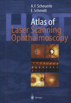
Atlas of Laser Scanning Ophthalmoscopy
Autor Alexander Friedrich Scheuerle Cuvânt înainte de H.E. Völcker Autor Eckart Schmidt Cuvânt înainte de L.E. Pillunat, F.E. Kruseen Limba Engleză Paperback – 19 ian 2012
Diseases of the optic nerve head; different types and stages of glaucoma, non-glaucomatous neuropathy and papilledema; automated classification procedures for the detection of glaucoma; strategies for the interpretation of follow-up results in optic disc monitoring.
Macular diseases: the shortly released Retina Module expands the diagnostic spectrum of laser scanning ophthalmoscopy significantly, adding measuring and monitoring of diabetic and cystoid macular edema.
This atlas is the most comprehensive up-to-date reference of laser scanning ophthalmoscopy available, ideal for residents and general ophthalmologists who want to enhance their diagnostic skills.
| Toate formatele și edițiile | Preț | Express |
|---|---|---|
| Paperback (1) | 339.08 lei 38-44 zile | |
| Springer Berlin, Heidelberg – 19 ian 2012 | 339.08 lei 38-44 zile | |
| Hardback (1) | 733.09 lei 6-8 săpt. | |
| Springer Berlin, Heidelberg – 3 dec 2003 | 733.09 lei 6-8 săpt. |
Preț: 339.08 lei
Preț vechi: 356.93 lei
-5% Nou
Puncte Express: 509
Preț estimativ în valută:
64.90€ • 70.52$ • 54.55£
64.90€ • 70.52$ • 54.55£
Carte tipărită la comandă
Livrare economică 17-23 aprilie
Preluare comenzi: 021 569.72.76
Specificații
ISBN-13: 9783642639210
ISBN-10: 3642639216
Pagini: 184
Ilustrații: XII, 170 p.
Dimensiuni: 193 x 260 x 10 mm
Ediția:2004
Editura: Springer Berlin, Heidelberg
Colecția Springer
Locul publicării:Berlin, Heidelberg, Germany
ISBN-10: 3642639216
Pagini: 184
Ilustrații: XII, 170 p.
Dimensiuni: 193 x 260 x 10 mm
Ediția:2004
Editura: Springer Berlin, Heidelberg
Colecția Springer
Locul publicării:Berlin, Heidelberg, Germany
V-ar putea interesa
-
 GlaucomaDeepak P. Edward-14%Preț: 577.14 lei672.50 lei
 -
 Uveitis: Fundamentals and Clinical Practice: Expert Consult - Online and PrintRobert B. Nussenblatt-25%Preț: 828.81 lei1112.36 lei
 -
 Thyroid Eye DiseaseRaymond S. Douglas-5%Preț: 655.15 lei689.63 lei
 -
 -22%Preț: 43.97 lei56.19 lei
 -
 -5%Preț: 572.97 lei603.13 lei
 -
 -5%Preț: 664.77 lei699.75 lei
 -
 -5%Preț: 433.46 lei456.28 lei
 -
 Dry Eye: A Practical ApproachColin Chan-5%Preț: 775.73 lei816.55 lei
 -
 Lens Epithelium and Posterior Capsular OpacificationShizuya Saika-5%Preț: 730.92 lei769.39 lei
 -
 -5%Preț: 1105.21 lei1163.38 lei
Public țintă
Professional/practitionerCuprins
1 Introduction.- 1.1 Why Is Laser Scanning Ophthalmoscopy Needed?.- 1.2 Technical Background.- 1.3 Stereometric Parameters of the HRT.- 2 Healthy Optic Discs.- 2.1 Natural Variety of Optic Discs.- 2.2 Regular Shapes.- 2.3 Micropapillas.- 2.4 Megalopapillas.- 2.5 Tilted Discs.- 3 Glaucomas.- 3.1 Automated Classification Procedures.- 3.2 Moderate Glaucomas.- 3.3 Nerve Fiber Bundle Defects.- 3.4 Advanced Glaucomas.- 3.5 Micropapillary Glaucomas.- 3.6 Megalopapillary Glaucomas.- 3.7 Normal Tension Glaucomas.- 3.8 Classification Errors.- 4 Follow-up Examinations.- 4.1 Strategies for Longitudinal Analysis.- 4.2 Progression of Glaucoma.- 5 Prominent Optic Discs.- 5.1 Scanning of Prominent Discs.- 5.2 Drusen of the Optic Nerve.- 5.3 Optic Nerve Swelling.- 5.4 Papilledemas.- 6 Macular Scans.- 6.1 The Macular Edema Software Module.- 6.2 Cystoid Macular Edema.- 6.3 Branch Vein Occlusion.- 6.4 Central Serous Retinopathy.- 6.5 Macular Puckers.- 6.6 Macular Holes.- 7 Miscellaneous.- 7.1 Optimizing Imaging.- 7.2 Astigmatism.- 7.3 Retinal Irregularities.- 7.4 Floaters.- 8 Confocal Corneal Imaging.- 8.1 The Rostock Cornea Modu.- References.
Textul de pe ultima copertă
Glaucoma remains one of the leading causes of blindness. Laser scanning tomography has gained an indispensable role in the ophthalmologic diagnosis, especially in the long-term follow-up of glaucoma. Confocal laser scanning ophthalmoscopy provides key insights into the three-dimensional anatomy of the optic disc in vivo. This unique atlas contains superb images of all clinically relevant diseases diagnosed by current models of the Heidelberg Retina Tomograph. It correlates classical diagnostic tools like perimetry, tonometry and fundus photography with state of the art studies including digital retinal angiography, optical coherence tomography and laser scanning tomography. Special features include the illustrated coverage of diseases of the optic nerve head; different types and stages of glaucoma, non-glaucomatous neuropathy and papilledema; automated classification procedures for the detection of glaucoma; strategies for the interpretation of follow-up results in optic disc monitoring and macular diseases. The shortly released Macular Edema Module expands the diagnostic spectrum of laser scanning ophthalmoscopy significantly, adding measuring and monitoring of diabetic and cystoid macular edema. This atlas is the most comprehensive up-to-date reference of laser scanning ophthalmoscopy available, ideal for residents and general ophthalmologists who want to enhance their diagnostic skills.
Caracteristici
First atlas for interpreting Heidelberg Retina Tomograph pictures Excellent teaching material Includes supplementary material: sn.pub/extras