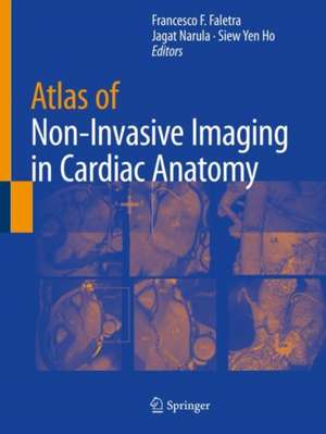
Atlas of Non-Invasive Imaging in Cardiac Anatomy
Editat de Francesco F. Faletra, Jagat Narula, Siew Yen Hoen Limba Engleză Paperback – 31 ian 2021
Atlas of Non-Invasive Imaging in Cardiac Anatomy provides a detailed set of visual instructions that is of use to any cardiovascular professional needing to understand the orientation of a patient’s imaging. Therefore this is an essential guide for all trainee and practicing cardiologists, cardiac imagers, cardiac surgeons and interventionists.
| Toate formatele și edițiile | Preț | Express |
|---|---|---|
| Paperback (1) | 652.02 lei 38-45 zile | |
| Springer International Publishing – 31 ian 2021 | 652.02 lei 38-45 zile | |
| Hardback (1) | 925.02 lei 38-45 zile | |
| Springer International Publishing – 31 ian 2020 | 925.02 lei 38-45 zile |
Preț: 652.02 lei
Preț vechi: 686.33 lei
-5% Nou
Puncte Express: 978
Preț estimativ în valută:
124.76€ • 130.26$ • 103.26£
124.76€ • 130.26$ • 103.26£
Carte tipărită la comandă
Livrare economică 31 martie-07 aprilie
Preluare comenzi: 021 569.72.76
Specificații
ISBN-13: 9783030355081
ISBN-10: 303035508X
Pagini: 133
Ilustrații: XI, 133 p. 9 illus.
Dimensiuni: 210 x 279 mm
Greutate: 0.45 kg
Ediția:1st ed. 2020
Editura: Springer International Publishing
Colecția Springer
Locul publicării:Cham, Switzerland
ISBN-10: 303035508X
Pagini: 133
Ilustrații: XI, 133 p. 9 illus.
Dimensiuni: 210 x 279 mm
Greutate: 0.45 kg
Ediția:1st ed. 2020
Editura: Springer International Publishing
Colecția Springer
Locul publicării:Cham, Switzerland
Cuprins
The Mitral Valve.- The Aortic Root.- The Tricuspid Valve.- The Interatrial Septum and the Septal Atrio-ventricular Junction.- The Right and the Left Atrium.- The Left and Right Ventricles.- The Coronary Arteries and Veins.
Recenzii
Notă biografică
Francesco Faletra, has been the head of the cardiac imaging department at Cardiocentro Ticino in Lugano, Switzerland since 2005. His focus is on 3D TEE and he regularly takes classes on this topic at cardiologists coming from all Europe. During his career, he has been the author of numerous books and chapters.
Jagat Narula is a world-renowned physician-scientist in cardiovascular medicine and imaging, and leads the cardiac services at Mount Sinai St. Luke’s Hospital. He is involved in clinical and basic research in the field of cardiovascular imaging. He has made contributions to the imaging of apoptotic cell death in heart muscle, and to the imaging of atherosclerotic plaques vulnerable to rupture.
S. Yen Ho is a Consultant Cardiac Morphologist with an international reputation for research and education in congenital heart disease and arrhythmias, including morphological correlates with cardiac imaging, surgery, interventions and arrhythmias. She organizes and directs numerous short courses on cardiac anatomy and congenital heart malformations for postgraduates and practitioners including the original and renowned ‘Hands-on Cardiac Morphology’ course at the Royal Brompton Hospital in London.
Textul de pe ultima copertă
This atlas provides a detailed visual resource of how sophisticated non-invasive imaging relates to the anatomy observed in a variety of cardiovascular pathologies. It includes investigation of a wide range of defects in numerous cardiac structures. Mitral valve commissures, atrioventricular septal junction and right ventricular outflow tract plus a wealth of other structures are covered, offering readers a comprehensive integrative experience to understand how anatomic subtleties are revealed by modern imaging modalities.
Atlas of Non-Invasive Imaging in Cardiac Anatomy provides a detailed set of visual instructions that is of use to any cardiovascular professional needing to understand the orientation of a patient’s imaging. Therefore this is an essential guide for all trainee and practicing cardiologists, cardiac imagers, cardiac surgeons and interventionists.
Atlas of Non-Invasive Imaging in Cardiac Anatomy provides a detailed set of visual instructions that is of use to any cardiovascular professional needing to understand the orientation of a patient’s imaging. Therefore this is an essential guide for all trainee and practicing cardiologists, cardiac imagers, cardiac surgeons and interventionists.
Caracteristici
Compares cardiovascular anatomy with exquisite imaging details provided by echo, CT and MRI Focuses on the anatomic structures that are most relevant for clinical cardiologists and cardiac interventionalists Provides a comprehensive integrative experience to understand the how imaging reflects anatomic subtleties
