Atlas of Ocular Optical Coherence Tomography
Editat de Fedra Hajizadehen Limba Engleză Hardback – 7 feb 2018
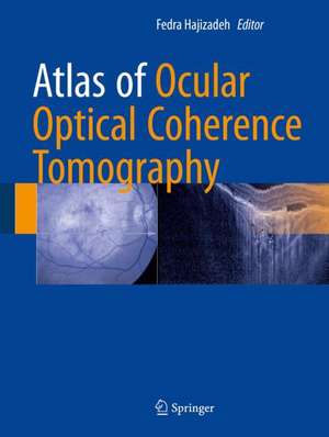
Atlas of Ocular Optical Coherence Tomography has been categorized into eleven sections, discussing and illustrating distinct OCT features, as well as showing other image modalities such as fluorescein angiography, fundus autofluorescence, perimetry and laboratory examination. This book also covers choroidal pathologies and vitreous abnormalities. The last section has been allocated to anterior segment disease, including cornea, angle, iris and conjunctival abnormalities. Above all, the numerous images, and detailed descriptions of diseases, make this book an essential guide for general ophthalmologists and ophthalmology residences.
| Toate formatele și edițiile | Preț | Express |
|---|---|---|
| Paperback (2) | 1189.85 lei 39-44 zile | |
| Springer International Publishing – 4 iun 2019 | 1189.85 lei 39-44 zile | |
| Springer International Publishing – 4 ian 2024 | 1560.00 lei 39-44 zile | |
| Hardback (2) | 590.43 lei 3-5 săpt. | +88.51 lei 7-13 zile |
| Springer International Publishing – 7 feb 2018 | 590.43 lei 3-5 săpt. | +88.51 lei 7-13 zile |
| Springer International Publishing – 3 ian 2023 | 2470.87 lei 3-5 săpt. |
Preț: 590.43 lei
Preț vechi: 621.50 lei
-5% Nou
Puncte Express: 886
Preț estimativ în valută:
113.01€ • 117.53$ • 94.70£
113.01€ • 117.53$ • 94.70£
Carte disponibilă
Livrare economică 20 februarie-06 martie
Livrare express 06-12 februarie pentru 98.50 lei
Preluare comenzi: 021 569.72.76
Specificații
ISBN-13: 9783319667560
ISBN-10: 3319667564
Pagini: 322
Ilustrații: IX, 483 p. 619 illus., 571 illus. in color.
Dimensiuni: 210 x 279 x 25 mm
Greutate: 2 kg
Ediția:1st ed. 2018
Editura: Springer International Publishing
Colecția Springer
Locul publicării:Cham, Switzerland
ISBN-10: 3319667564
Pagini: 322
Ilustrații: IX, 483 p. 619 illus., 571 illus. in color.
Dimensiuni: 210 x 279 x 25 mm
Greutate: 2 kg
Ediția:1st ed. 2018
Editura: Springer International Publishing
Colecția Springer
Locul publicării:Cham, Switzerland
Cuprins
Foreword.- Introduction.- Age related macular degeneration (ARMD) .- Diabetic retinopathy and retinal vascular diseases.- Central serous chorioretinopathy (CSCR).- Epiretinal membrane, macular hole and vitreomacular traction (VMT) syndrome..- Optic disc anomalies .- Ocular tumors.- Pathologic Myopia.- Hereditary disease.- Uveitis and intraocular inflammation.- Anterior segment OCT.
Notă biografică
Fedra Hajizadeh, MD has been a Vitreoretinal Consultant and Ophthalmology Surgeon at Noor Eye Hospital, Tehran, Iran, since March 2008. Whilst working there she has also been in charge of the of OCT and angiography departments. Prior to her work at Noor Eye Hospital, she has had experience as a surgeon and consultant at Chamran Hospital, Farabi Hospital, and Firooz Abadi Hospital. She has undertaken years of research, securing a number of certificates and honours such as a second place for “Optic Pit Related Foveoschisis” at the Ophthalmic Photographers Society Exhibit, ASCRS-ASOA Symposium & Congress, Boston, Massachusetts, USA. She has published research in numerous local and international journals.
Textul de pe ultima copertă
This book provides a collection of optical coherence tomographic (OCT) images of various diseases of posterior and anterior segments. It covers the details and issues of diagnostic tests based on OCT findings which are crucial for ophthalmologists to understand in their clinical practice. Throughout the chapters all aspects of this non-invasive, popular imaging technique, known for ingenuity and accuracy, is clearly illustrated.
Atlas of Ocular Optical Coherence Tomography has been categorized into eleven sections, discussing and illustrating distinct OCT features, as well as showing other image modalities such as fluorescein angiography, fundus autofluorescence, perimetry and laboratory examination. This book also covers choroidal pathologies and vitreous abnormalities. The last section has been allocated to anterior segment disease, including cornea, angle, iris and conjunctival abnormalities. Above all, the numerous images, and detailed descriptions of diseases, make this book an essential guide for general ophthalmologists and ophthalmology residences.
Atlas of Ocular Optical Coherence Tomography has been categorized into eleven sections, discussing and illustrating distinct OCT features, as well as showing other image modalities such as fluorescein angiography, fundus autofluorescence, perimetry and laboratory examination. This book also covers choroidal pathologies and vitreous abnormalities. The last section has been allocated to anterior segment disease, including cornea, angle, iris and conjunctival abnormalities. Above all, the numerous images, and detailed descriptions of diseases, make this book an essential guide for general ophthalmologists and ophthalmology residences.
Caracteristici
Unparalleled scope and range of images
Detailed descriptions of diseases
Covers the pitfalls and issues of OCT use
Detailed descriptions of diseases
Covers the pitfalls and issues of OCT use
