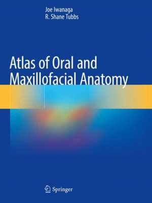
Atlas of Oral and Maxillofacial Anatomy
Autor Joe Iwanaga, R. Shane Tubbsen Limba Engleză Paperback – 6 noi 2022
| Toate formatele și edițiile | Preț | Express |
|---|---|---|
| Paperback (1) | 770.61 lei 38-44 zile | |
| Springer International Publishing – 6 noi 2022 | 770.61 lei 38-44 zile | |
| Hardback (1) | 1040.25 lei 38-44 zile | |
| Springer International Publishing – 6 noi 2021 | 1040.25 lei 38-44 zile |
Preț: 770.61 lei
Preț vechi: 811.17 lei
-5% Nou
Puncte Express: 1156
Preț estimativ în valută:
147.45€ • 154.37$ • 122.01£
147.45€ • 154.37$ • 122.01£
Carte tipărită la comandă
Livrare economică 01-07 aprilie
Preluare comenzi: 021 569.72.76
Specificații
ISBN-13: 9783030783297
ISBN-10: 3030783294
Pagini: 168
Ilustrații: VII, 168 p. 358 illus., 342 illus. in color.
Dimensiuni: 210 x 279 mm
Ediția:1st ed. 2021
Editura: Springer International Publishing
Colecția Springer
Locul publicării:Cham, Switzerland
ISBN-10: 3030783294
Pagini: 168
Ilustrații: VII, 168 p. 358 illus., 342 illus. in color.
Dimensiuni: 210 x 279 mm
Ediția:1st ed. 2021
Editura: Springer International Publishing
Colecția Springer
Locul publicării:Cham, Switzerland
Cuprins
Anatomy of the Superficial Face.- Bones of the Head and Neck.- Cranial Fossae and Related Structures.- Venous Drainage of the Oral and Maxillofacial Area.- Cranial Nerves and Spinal Nerves.- Anatomy of the Oral Cavity.- Maxillofacial Anatomy.- Spaces and Fossa in the Oral and Maxillofacial Area.
Notă biografică
Joe Iwanaga, DDS, PhD, is an Associate Professor in the Department of Neurosurgery at Tulane University School of Medicine, New Orleans, LA, USA . He is also an Associate Professor in the Department of Anatomy at Kurume University School of Medicine, Kurume, Fukuoka, Japan. Dr. Iwanaga graduated from Tokyo Medical and Dental University, School of Dentistry. He has expertise in both oral and maxillofacial surgery and clinical anatomy, and his research and surgical focus is on anatomical variations and microsurgical anatomy. He has delivered numerous presentations and is the author of more than 400 articles in peer-reviewed journals. Dr. Iwanaga is a Councilor-at-Large of the American Association of Clinical Anatomists and in 2017–8 was Presidential Appointee of the Nominating Committee for the American Association of Clinical Anatomists. He is a Dental, Oral and Maxillofacial Section editor of the journal Clinical Anatomy. He also acted as an international advisory board of the 7th edition of Netter’s Atlas of Human Anatomy and authored 42nd edition of Gray’s Anatomy (Elsevier).
R. Shane Tubbs, MS, PA-C, PhD is a clinical anatomist, author, editor, and researcher. He is Professor of Neurosurgery, Neurology, Surgery, and Structural & Cellular Biology, Director of Surgical Anatomy at Tulane School of Medicine and Program Director of Anatomical Research in the Clinical Neuroscience Research Center at Tulane University School of Medicine, New Orleans, Louisiana. He has honorary professorships/faculty positions at St. George’s University, Grenada, University of Queensland, Brisbane, Australia, Department of Neurosurgery, Ochsner Medical Center, New Orleans, Louisiana, and the National Skull Base Center of California. He is Editor-in-Chief of the journal Clinical Anatomy and Chair of the Federative International Programme on Anatomical Terminologies (FIPAT). Dr. Tubbs’ research interests are centered around what has been termed “reverse translational anatomy research” where clinical/surgical problems are identified and solved/explained with anatomical studies. This investigative paradigm in anatomy has resulted in over 1,700 peer reviewed publications from his laboratory. His h-index is 72 and in 2018, he was listed as a “hyperprolific author” in the journal Nature.
Dr. Tubbs has authored/edited over 50 books and over 80 book chapters primarily in anatomical and neurosurgical textbooks. His textbooks, include the first and second editions of The Chiari Malformations, Gray’s Anatomy Review editions 1-3, Gray’s Clinical Photographic Dissector of the Human Body editions 1-3, Netter’s Introduction to Clinical Procedures, Anatomy for Plastic Surgery of the Face, Head, and Neck, Nerves and Nerve Injuries volumes I and II, Occult Spinal Dysraphism, An Illustrated Terminologia Neuroanatomica, Surgical Anatomyof the Lumbar Plexus, A History of Human Anatomy, A Guide to the Scientific Career, Kerr’s Brachial Plexus, Hamilton’s History of Medicine and Surgery, Surgical Anatomy of the Lateral Transpsoas Approach to the Lumbar Spine, Anatomy, Imaging, and Surgery of the Intracranial Dural Venous Sinuses, The Island of Reil (Insula) in the Human Brain, and Bergman’s Comprehensive Encyclopedia of Human Anatomic Variation. He is an editor for the 41st and 42nd editions of the over 150-year-old Gray’s Anatomy, and the 5th through 8th editions of Netter’s Atlas of Anatomy
Textul de pe ultima copertă
This comprehensive atlas, featuring a wealth of top quality photographs of fresh cadaveric dissections, is a superb guide to anatomic structures in the oral and maxillofacial region that will be an ideal aid in clinical practice. It has the important benefit of enabling readers to observe the anatomy from the same view as seen during invasive clinical procedures. This is critical for a better understanding of these procedures, and surgical annotations are included as necessary. Atlas of Oral and Maxillofacial Anatomy is the first book of its kind to be devoted to the clinical anatomy of the region for dentists and oral and maxillofacial surgeons. It will satisfy the demand for such a comprehensive atlas in this field of surgery and will be welcome and timely for clinicians and trainees. Beyond specialists and residents in oral and maxillofacial surgery and general dentists, the book will be of value for craniofacial surgeons, anatomists, plastic surgeons, ENT surgeons, head andneck surgeons, neurosurgeons, dental students, medical students, dental hygienists, and nurses working with dentists and oral and maxillofacial surgeons.
Caracteristici
Top quality photographs of fresh cadaveric dissections Anatomy shown as seen during invasive clinical procedures Ideal aid for dentists and oral and maxillofacial surgeons
