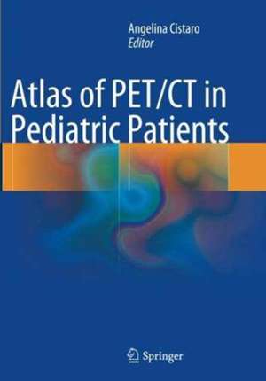
Atlas of PET/CT in Pediatric Patients
Editat de Angelina Cistaroen Limba Engleză Paperback – 2 oct 2016
| Toate formatele și edițiile | Preț | Express |
|---|---|---|
| Paperback (1) | 898.67 lei 38-44 zile | |
| Springer – 2 oct 2016 | 898.67 lei 38-44 zile | |
| Hardback (1) | 729.98 lei 3-5 săpt. | |
| Springer – 26 noi 2013 | 729.98 lei 3-5 săpt. |
Preț: 898.67 lei
Preț vechi: 945.98 lei
-5% Nou
Puncte Express: 1348
Preț estimativ în valută:
172.01€ • 186.91$ • 144.59£
172.01€ • 186.91$ • 144.59£
Carte tipărită la comandă
Livrare economică 17-23 aprilie
Preluare comenzi: 021 569.72.76
Specificații
ISBN-13: 9788847058729
ISBN-10: 8847058724
Pagini: 264
Ilustrații: XII, 264 p. 181 illus., 170 illus. in color.
Dimensiuni: 178 x 254 mm
Ediția:Softcover reprint of the original 1st ed. 2014
Editura: Springer
Colecția Springer
Locul publicării:Milano, Italy
ISBN-10: 8847058724
Pagini: 264
Ilustrații: XII, 264 p. 181 illus., 170 illus. in color.
Dimensiuni: 178 x 254 mm
Ediția:Softcover reprint of the original 1st ed. 2014
Editura: Springer
Colecția Springer
Locul publicării:Milano, Italy
Cuprins
Section I Basic Science and Practical Issues 1 Radiofarmaceutical Compounds.- 2 Method and Preparation of the Patient.- 3 18F-FDG Administration and Dosimetry.- 4 Physiological Pattern and Pitfall of 18F-FDG Biodistribution.- Section II Oncology 5 Malignant Lymphoma in Children.- 6 18-FDG PET/CT in Pediatric Lymphoma.- 7 Other Haematological Diseases (Leukemia).- 8 Primary Bone Tumors.- 9 Utility of 18F-FDG PET/CT in Soft Tissue Sarcomas.- 10 Primary Hepatic Tumors.- 11 Neuroendocrine Tumors.- 12 Neuroblastoma.- Other Tumors 13 Pediatric Nasopharyngeal Carcinoma.- 14 Poorly Differentiated Thyroid Carcinoma.- 15 Phylloid Tumor of the Breast.- 16 Wilms Tumor.- 17 Adrenal Gland Cancer.- 18 Ovarian Teratoma.- Section III Neurology 19 Role of Aminoacid PET Tracers in Pediatric Brain Tumors.- 20 18F-FDG in Brain Tumors.- 21 PET/CT in the Clinical Evaluation of Pediatric Epilepsy.- 22 Epilepsia Partialis Continua.- 23 Brain 18F- FDG PET/CT Imaging in Haemolytic Uraemic Syndrome (HUS) During and After the Acute Phase.- Section IV Infection and Inflammation 24 Inflammatory Bowel Disease.- 25 Appendicitis.- 26 Spondylodiscitis.- 27 Other Bone Lesion.- 28 Pulmonary Aspergillosis.- 29 Mycobatteriosis.- Section V Other Applications 30 Sarcoidosis.- 31 Neurofibromatosis.- 32 Autoimmune Lymphoproliferative Syndrome (ALPS). 33 Castelman’s Disease.- 34 Fever of Unknown Origin.- 35 Congenital Hyperinsulinism.- 36 Myocardial Perfusion Imaging with Rb Cardiac PET/CT.
Recenzii
From the book reviews:
“The book is well written and highly didactic, therefore having a very high appeal for residents and practitioners, first of all for those involved in pediatrics. In my opinion, this volume has to be present in all nuclear medicine labs and is also useful for nuclear physicians, radiologists, oncologists, clinicians, surgeons, and all people who want to be updated on the state of the art and on clinical indications for PET/CT in infants and young patients.” (Luigi Mansi, European Journal of Nuclear Medicine and Molecular Imaging, Vol. 41, 2014)
“The book is well written and highly didactic, therefore having a very high appeal for residents and practitioners, first of all for those involved in pediatrics. In my opinion, this volume has to be present in all nuclear medicine labs and is also useful for nuclear physicians, radiologists, oncologists, clinicians, surgeons, and all people who want to be updated on the state of the art and on clinical indications for PET/CT in infants and young patients.” (Luigi Mansi, European Journal of Nuclear Medicine and Molecular Imaging, Vol. 41, 2014)
Textul de pe ultima copertă
This richly illustrated book presents the pediatric applications of PET/CT in the full range of scenarios frequently encountered in a professional setting. It opens with a thorough introduction covering the fundamental science and the clinical basis for use of PET/CT in this age group. Pitfalls and artifacts are examined, and normal variations and benign findings are carefully described. Each subsequent chapter addresses the role of PET/CT with different radiopharmaceuticals in the evaluation and management of a specific disease. The full range of oncological diseases is covered, including the rare ones. Succinct descriptions of clinical cases are included and, when appropriate, comparisons are made with other modalities. In addition, the role of PET/CT in biopsy guidance and in radiation therapy planning is explained. This book will be invaluable for residents and practitioners in nuclear medicine, radiology, oncology, radiation oncology, and nuclear medicine technology
Caracteristici
Numerous PET/CT images of various pediatric tumors PET/CT images of rare pediatric pathologies Identification of pitfalls and artifacts Guidance on the optimal performance of PET/CT scans
