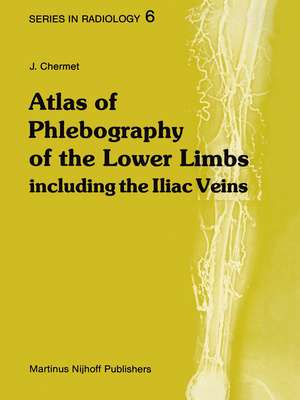
Atlas of Phlebography of the Lower Limbs: Including the Iliac Veins: Series in Radiology, cartea 6
Autor J. Chermeten Limba Engleză Paperback – 9 oct 2011
Din seria Series in Radiology
- 5%
 Preț: 437.14 lei
 - 5%
 Preț: 370.21 lei
 - 5%
 Preț: 366.19 lei
 - 5%
 Preț: 348.95 lei
 - 5%
 Preț: 366.19 lei
 -
 Preț: 367.86 lei
 - 5%
 Preț: 369.45 lei
 - 5%
 Preț: 363.60 lei
 - 5%
 Preț: 396.71 lei
 - 5%
 Preț: 366.19 lei
 - 5%
 Preț: 340.64 lei
 - 5%
 Preț: 715.91 lei
 - 5%
 Preț: 710.06 lei
 - 5%
 Preț: 367.07 lei
 - 5%
 Preț: 715.91 lei
 - 5%
 Preț: 385.94 lei
 - 5%
 Preț: 1408.43 lei
 - 5%
 Preț: 670.95 lei
 - 5%
 Preț: 1416.30 lei
Preț: 379.69 lei
Preț vechi: 399.67 lei
-5% Nou
Puncte Express: 570
Preț estimativ în valută:
72.65€ • 76.06$ • 60.12£
72.65€ • 76.06$ • 60.12£
Carte tipărită la comandă
Livrare economică 07-21 aprilie
Preluare comenzi: 021 569.72.76
Specificații
ISBN-13: 9789400974630
ISBN-10: 9400974639
Pagini: 304
Ilustrații: VII, 294 p.
Dimensiuni: 210 x 279 x 16 mm
Greutate: 0.69 kg
Ediția:Softcover reprint of the original 1st ed. 1982
Editura: SPRINGER NETHERLANDS
Colecția Springer
Seria Series in Radiology
Locul publicării:Dordrecht, Netherlands
ISBN-10: 9400974639
Pagini: 304
Ilustrații: VII, 294 p.
Dimensiuni: 210 x 279 x 16 mm
Greutate: 0.69 kg
Ediția:Softcover reprint of the original 1st ed. 1982
Editura: SPRINGER NETHERLANDS
Colecția Springer
Seria Series in Radiology
Locul publicării:Dordrecht, Netherlands
Public țintă
ResearchCuprins
1. Radioanatomy and venographic illustration of the techniques. Figures 1–62.- 2. Artefacts and incidents. Figures 63–100.- 3. Acute thrombophlebitis of the veins of the lower limb. Figures 101–127..- 4. Iliofemoral and iliocaval phlebites. Thrombosis of the inferior vena cava. Figures 128–167.- 5. Collateral circulation in case of obliteration of the veins of the lower limbs and the iliac veins. Figures 168–203.- 6. Surgery in iliofemoral phlebites. Figures 204–224.- 7. Chronic phlebitis and surgery in chronic phlebitis. Figures 225–285.- 8. Varices and venography. Figures 286–304.- 9. Recurrent varicose veins. Operative treatment of varicose veins. Figures 305–316.- 10. Acquired and congenital valvular incompetence. Femoral and popliteal retrograde venography. Figures 317–331.- 11. Extrinsic compression of the veins of the lower limb and of the iliac veins. Cockett’s syndrome. Figures 332–374.- 12. Venous dysplasiae. Figures 375–397.- 13. Traumatism to the veins. Figures 398–404.- 14. Various pathologic conditions of the veins of the lower limb. Figures 405–409.- 15. Radioisotope phlebography, by Aslam R. Siddiqui, M.D. (Indiana University Medical Center, Division of Nuclear Medicine, Indianapolis). Figures 410–418.
Recenzii
`The quality of the illustrations is quite good, and it is evident that the author knows his business. The figures are labeled very clear, and labeled structures in each illustration can be identified rapidly, with only a glance at the accompanying legend. ...it will be of value to those with a basic knowledge of venography, who are likely to profit from its excellent illustrations. It deserves to be in any department where venography is a common procedure, and in the library of every residence program, as a supplement to textbooks on venography.'
American Journal of Roentgenology
`It gives me great pleasure to review this atlas by Professor Chermet which is part of a series of radiology atlases intended to cover the main aspects of medical imaging. ... The illustrations are almost universally excellent examples of the conditions they represent, and very well produced, and for this alone the author and publishers are to be congratulated. ... I strongly recommend this atlas, which, as far as I am aware, is the only one available in the English language and which, considering the large number of first-class illustrations, is by today's standards really quite cheap. I hope it will be available in every radiology department where phlebograms are, even if only occasionally requested.'
British Journal of Radiology
