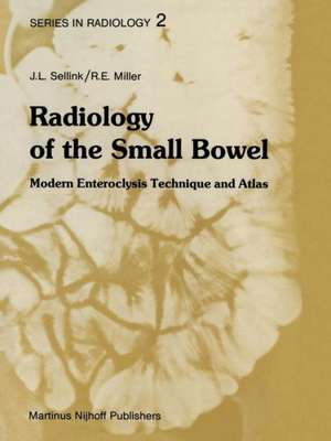
Radiology of the Small Bowel: Modern Enteroclysis Technique and Atlas: Series in Radiology, cartea 2
Autor J.L. Sellink, D.J. Milleren Limba Engleză Paperback – 8 oct 2011
Din seria Series in Radiology
- 5%
 Preț: 437.14 lei
 - 5%
 Preț: 370.21 lei
 - 5%
 Preț: 366.19 lei
 - 5%
 Preț: 348.95 lei
 - 5%
 Preț: 366.19 lei
 -
 Preț: 367.86 lei
 - 5%
 Preț: 369.45 lei
 - 5%
 Preț: 363.60 lei
 - 5%
 Preț: 366.19 lei
 - 5%
 Preț: 340.64 lei
 - 5%
 Preț: 715.91 lei
 - 5%
 Preț: 710.06 lei
 - 5%
 Preț: 367.07 lei
 - 5%
 Preț: 715.91 lei
 - 5%
 Preț: 385.94 lei
 - 5%
 Preț: 1408.43 lei
 - 5%
 Preț: 379.69 lei
 - 5%
 Preț: 670.96 lei
 - 5%
 Preț: 1416.30 lei
Preț: 396.71 lei
Preț vechi: 417.59 lei
-5% Nou
Puncte Express: 595
Preț estimativ în valută:
75.92€ • 78.97$ • 62.68£
75.92€ • 78.97$ • 62.68£
Carte tipărită la comandă
Livrare economică 15-29 aprilie
Preluare comenzi: 021 569.72.76
Specificații
ISBN-13: 9789400974326
ISBN-10: 9400974329
Pagini: 500
Ilustrații: 495 p.
Dimensiuni: 210 x 280 x 26 mm
Greutate: 1.11 kg
Ediția:Softcover reprint of the original 1st ed. 1982
Editura: SPRINGER NETHERLANDS
Colecția Springer
Seria Series in Radiology
Locul publicării:Dordrecht, Netherlands
ISBN-10: 9400974329
Pagini: 500
Ilustrații: 495 p.
Dimensiuni: 210 x 280 x 26 mm
Greutate: 1.11 kg
Ediția:Softcover reprint of the original 1st ed. 1982
Editura: SPRINGER NETHERLANDS
Colecția Springer
Seria Series in Radiology
Locul publicării:Dordrecht, Netherlands
Public țintă
ResearchCuprins
1. Introduction.- 2. Anatomy.- 1. Normal mucosa in the small intestine.- 2. Normal position of the intestine.- 3. Normal impressions on the intestine.- 4. Filling defects between the intestinal loops.- 3. Physiology.- 1. Innervation and motility.- 2. Gastric emptying and transit time.- 4. The Contrast Medium.- 1. General considerations.- 2. Sedimentation of the contrast medium.- 3. Flocculation of the contrast fluid.- 4. Segmentation of the contrast column.- 5. Additives to the contrast medium for the purpose of improving stability and adhesion.- 6. Relationship between viscosity, particle size, and adhesion of the barium suspension.- 7. Specific gravity of the contrast fluid.- 8. Contrast media other than barium sulfate.- 5. Methods of Examination.- 1. ‘Physiological’ examination of the small intestine.- 2. Single administration of the contrast medium.- 3. Fractional administration of the contrast medium.- 4. Administration of cold fluids with the contrast medium.- 5. Administration of the contrast medium through a tube directly into the small intestine (enteroclysis).- 6. Retrograde administration of the contrast fluid.- 7. Combined methods of examination.- 8. Use of drugs to accelerate transit.- 6. General Considerations.- 7. The Enteral Contrast Infusion.- 1. Preparation of patients.- 2. Duodenal intubation.- 3. Partial gastrectomy.- 4. Special types of tubes.- 5. Administration of contrast fluid.- 6. Administration of water after the barium suspension.- 7. Administration of air after contrast fluid.- 8. Compression technique.- 8. Basic Signs of Abnormality.- 1. Changes in the mucosal patterns.- 2. Lymph follicles — nodules — polyps.- 3. Foreign bodies and filling defects in the contrast fluid.- 4. Ulcerations.- 5. Deformation of the intestine.- 6. Dilutionof the contrast fluid — haziness — mucus secretion.- 7. Disintegration and misleading patterns.- 8. Malabsorption.- 9. Motility 180 Bibliography: chapters 1–8.- 9. Inflammation and Inflammatory-Like Diseases.- 1. General.- 2. Crohn’s disease.- 3. Reflux ileitis.- 4. Yersinia EC infections.- 5. Eosinophilic gastroenteritis.- 6. Radiation enteritis.- 7. Whipple’s disease.- 8. Aspecific ulcers.- 9. Appendicular infiltrates.- 10. Zollinger-Ellison disease.- 11. Radiological manifestations of serum protein disorders.- 10. Tumors.- 1. General.- 2. Polyposis.- 3. Benign tumors.- 4. Semimalignant tumors.- 5. Malignant tumors.- 6. Metastasis.- 11. Vascular Diseases.- 1. Ischemia due to impaired arterial flow.- 2. Graft versus host syndrome.- 3. Impaired venous flow.- 4. Periodic vascular insufficiency.- 5. Hemorrhage.- 12. Disturbed Motility.- 1. Drug-induced atony of the small bowel.- 2. Collagen diseases.- 3. Adult celiac disease (W.F.H. Müller).- 4. Amyloidosis.- 13. Congenital Anomalies.- 1. Abnormal positioning of the entire small bowel: disturbed rotation or fixation.- 2. Abnormal or fixed positioning of several intestinal loops: internal hernia.- 3. Duplications.- 4. Diverticulosis.- 5. Meckel’s diverticulum.- 14. Ileus — Fusion — Bands — Volvulus — Intussusceptions — Incisional Hernia.- 1. Ileus.- 2. Fusion — bands.- 3. Volvulus.- 4. Intussusceptions.- 5. Incisional hernia.- 15. The Enteroclysis Examination of Infants.- 1. Preparation.- 2. Duodenal intubation.- 3. The contrast fluid.- 4. The examination.- 5. Results.- 16. Common Errors and Failures.- 1. Preparation.- 2. Execution of the examination.- 3. General mistakes and failures.
Recenzii
`In summary, the high standard of radiology of the small bowel using modern enteroclysis technique and their result by Sellink and Miller is once again most impressive. This volume is an invaluable reference for radiologists and gastroenterologists.'
European Journal of Nuclear Medicine, 8:9 (1983)
`This is a cathedral of a book, full of detail, fussy, tiring, but in the end grand. ... The text is comprehensive and the book is lavishly illustrated. ... The chapter on technique is excellent and describes in detail the factors necessary for obtaining consistently good results. ... This book should stimulate radiologists to take a greater interest in the subject and a copy of the book should be available in all departmental libraries.'
The British Journal of Radiology, 56:669 (1983)
'Although entitled as an atlas, the book has a large amount of excellent material. The illustrations are of outstanding quality. ... This book is destined to become a classic in the radiologic literature and is highly recommended to all readers interested in diseases of the small bowel.'
The New England Journal of Medicine
`The authors and the publisher are to be congratulated for the outstanding illustrations with more than 1,000 uniformly superb radiographs. No radiograph less than excellent has been allowed... The text fully covers the complex subject and correlates well with the illustrations. ... Certainly the radiologist who wishes to offer comlete gastrointestinal radiology needs to include enteroclysis and needs to have this book.'
American Journal of Roentgenology
`This is quite a splendid atlas which will be indispensable to any radiologist wishing to explore this technique. It should beavailable to all trainees in radiology who are likely to be encouraged by the superb illustrations of a wide range of small bowel pathology to try out the method for themselves.'
Gut
`Radiology of the Small Bowel is a major contribution to a field which deserves more attention to detail and technique. ... The authors have produced an extremely well-organized and well-written atlas that contains a tremendous amount of beautifully illustrated pathologic material. The book succeeds in describing the proper technique of enteroclysis and in illustrating all conceivable abnormalities of the small bowel. The quality of the printing and illustrations is superior... This wonderfully written and illustrated atlas is a valuable contribution to the literature. ...recommended for general radiologists, gastrointestinal radiologists, and gastroenterologists.'
Radiology
`An excellent atlas of pathologic anatomy as demonstrated by enteroclysis. Almost all conceivable small bowel lesions are demonstrated and discussed. ...the illustrations are unusually good.'
Gastroenterology
