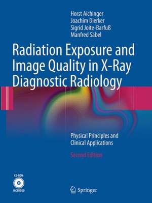
Radiation Exposure and Image Quality in X-Ray Diagnostic Radiology: Physical Principles and Clinical Applications
Autor Horst Aichinger, Joachim Dierker, Sigrid Joite-Barfuß, Manfred Säbelen Limba Engleză Paperback – 4 mai 2017
| Toate formatele și edițiile | Preț | Express |
|---|---|---|
| Paperback (1) | 578.37 lei 39-44 zile | |
| Springer Berlin, Heidelberg – 4 mai 2017 | 578.37 lei 39-44 zile | |
| Mixed media product (1) | 837.64 lei 3-5 săpt. | |
| Springer Berlin, Heidelberg – 22 noi 2011 | 837.64 lei 3-5 săpt. |
Preț: 578.37 lei
Preț vechi: 608.81 lei
-5% Nou
Puncte Express: 868
Preț estimativ în valută:
110.69€ • 116.78$ • 92.25£
110.69€ • 116.78$ • 92.25£
Carte tipărită la comandă
Livrare economică 30 decembrie 24 - 04 ianuarie 25
Preluare comenzi: 021 569.72.76
Specificații
ISBN-13: 9783662517543
ISBN-10: 366251754X
Pagini: 307
Ilustrații: XIV, 307 p. 229 illus.
Dimensiuni: 210 x 279 x 34 mm
Ediția:Softcover reprint of the original 2nd ed. 2012
Editura: Springer Berlin, Heidelberg
Colecția Springer
Locul publicării:Berlin, Heidelberg, Germany
ISBN-10: 366251754X
Pagini: 307
Ilustrații: XIV, 307 p. 229 illus.
Dimensiuni: 210 x 279 x 34 mm
Ediția:Softcover reprint of the original 2nd ed. 2012
Editura: Springer Berlin, Heidelberg
Colecția Springer
Locul publicării:Berlin, Heidelberg, Germany
Cuprins
Physical principles: Production and measurement of X-rays.- Interaction of photons with matter.- Radiation field and dosimetric quantities.- Penetration of X-rays.- Scattered radiation.- Image receptors.- Image quality and dose.- Clinical Applications: Evaluation of dose to the patient.- Scattered radiation.- Optimisation of image quality and dose.- Supplement: X-ray spectra.- Interaction coefficients.- Characteristics of the primary radiation beam.- Characteristics of the imaging radiation field.- Miscellaneous.- Patient-dose-estimation.
Recenzii
From the reviews:
“This well-written hardback describes the physical principles involved in imaging with x-rays. … an excellent addition to any medical physics department that has involvement in diagnostic radiology. It is suitable for trainee physicists in need of an up-to-date imaging reference, and will also be useful to experienced staff to refresh their knowledge on specific topics or make use of the reference data.” (David Platten, Scope, Vol. 22 (4), December, 2013)
"This textbook deals with the central concern of professionals working in diagnostic imaging. … it treats the material in greater depth than is usual in standard radiological physics textbooks. … The strength of the work resides in its structured treatment of topics that are pivotal in diagnostic imaging and also CT. … I would recommend it as an invaluable reference work for scientists working in diagnostic imaging. It should be of particular value to those involved in the training of staff in the field." (Dr M Casey, RAD Magazine, May, 2004)
"The book derives from a wide range of knowledge, scientific data, and extensive practical experience accumulated over the years. … This book, which is a clear and comprehensive scientific overview, provides a reference for physicists, engineers, and other experts working on problems of image quality, patient dose estimation, and the establishment of diagnostic reference levels. Its launch at this time is well chosen, as European experts are much concerned with the demands of CEC 97/43 publication (MED)." (O. Glomset, Acta Radiologica, 2004)
Textul de pe ultima copertă
Diagnostic X-rays are the largest contributor to radiation exposure to the general population, and protecting the patient from radiation damage is a major aim of modern health policy. Once the decision has been taken to use ionising radiation for imaging in a particular patient, it is necessary to optimize the image acquisition process taking into account the diagnostic quality of the images and the radiation dose to the patient. Both image quality and radiation dose are affected by a number of parameters, knowledge of which permits scientifically based decision making.
The authors of this second edition of Radiation Exposure and Image Quality in X-ray Diagnostic Radiology have spent many years studying the optimization of radiological imaging. In this book they present in detail the basic physical principles of diagnostic radiology and their application to clinical problems. Particular attention is devoted to evaluation of the dose to the patient, the influence of scattered radiation on image quality, the use of antiscatter grids, and optimization of image quality and dose. The final section is a supplement containing tables of data and graphical depictions of X-ray spectra, interaction coefficients, characteristics of X-ray beams, and other aspects relevant to patient dose calculations. In addition, a complementary CD-ROM contains a user-friendly Excel file database covering these aspects that can be used in the reader’s own programs.
Since the first edition, the text, figures, tables, and references have all been thoroughly updated, and more detailed attention is now paid to image quality and radiation exposure when using digital imaging and computed tomography. This book will be an invaluable aid to medical physicists when performing calculations relating to patient dose and image quality, and will also prove useful for diagnostic radiologists and engineers.
Caracteristici
Thoroughly updated new edition that examines in detail the relationship between radiation dose and image quality, including when using digital imaging and computed tomography Written by experts who for many years have collected data relevant to the optimization of radiological diagnosis Complementary CD-ROM containing a user-friendly Excel file that can be used in calculations
