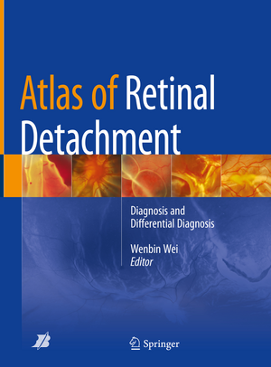
Atlas of Retinal Detachment: Diagnosis and Differential Diagnosis
Editat de Wenbin Weien Limba Engleză Hardback – 28 ian 2019
The book includes both common types of retinal detachment and some rare diseases. Moreover, some diseases without but related to retinal detachment are included in the book. In addition to traditional color fundus photos, fundus fluorescent angiography, ultrasound and traditional OCT images, OCT angiography and ultrasound angiography images are included too. This book also contains brief introductions to each disease. Differentiated from detailed review of pathogenesis or treatment, the readers will discover concise yet direct concept.
This atlas and textbook is yours to read and enjoy, but also to use as a valuable tool in your management of the patient with retinal detachment. Keep it handy. We hope doctors in training and those who have had certain clinical experiences interested in vitro-retinal diseases can benefit from this book.
Preț: 1509.12 lei
Preț vechi: 1588.56 lei
-5% Nou
Puncte Express: 2264
Preț estimativ în valută:
288.76€ • 302.31$ • 238.94£
288.76€ • 302.31$ • 238.94£
Carte tipărită la comandă
Livrare economică 02-08 aprilie
Preluare comenzi: 021 569.72.76
Specificații
ISBN-13: 9789811082306
ISBN-10: 9811082308
Pagini: 340
Ilustrații: XI, 262 p. 415 illus., 353 illus. in color.
Dimensiuni: 210 x 279 x 14 mm
Greutate: 1.26 kg
Ediția:1st ed. 2018
Editura: Springer Nature Singapore
Colecția Springer
Locul publicării:Singapore, Singapore
ISBN-10: 9811082308
Pagini: 340
Ilustrații: XI, 262 p. 415 illus., 353 illus. in color.
Dimensiuni: 210 x 279 x 14 mm
Greutate: 1.26 kg
Ediția:1st ed. 2018
Editura: Springer Nature Singapore
Colecția Springer
Locul publicării:Singapore, Singapore
Cuprins
Chapter 1: Introduction of Retinal Detachment (Ma Kai).- Chapter 2: Rhegmatogenous Retinal Detachment (Zhu Ruilin).- Chapter 3: Special Types of Rhegmatogenous Retinal Detachment (Ma Kai).- Chapter 4: Exudative Retinal Detachment (Zhou Haiying).- Chapter 5: Fundus Congenital Abnormalities and Retinal Detachment (Huang Yao).- Chapter 6: Tractional retinal detachment (Shao Lei).- Chapter 7: Traumatic Retinal Detachment (Zhou Jinqiong).- Chapter 8: Pathologic Myopia and Retina Detachment (Gao Liqin).- Chapter 9: Infectious Uveitis and Retinal Detachment (Gao Liqin).- Chapter 10: Uveitis and retinal detachment (Huang Yao).- Chapter 11: Ocular Tumor and Retinal Detachment.- Chapter 12: Surgical Techniques of Rhegmatogenous Retinal Detachment and Complications After Surgeries (She Haicheng).
Notă biografică
Wenbin Wei is a professor and the director of the Department of Ophthalmology, Beijing Tongren Hospital, Capital Medical University, Beijing, China.
Textul de pe ultima copertă
This atlas and textbook aims to provide diverse pictures, facilitating the readers to learn fundus diseases. Retinal detachment is a common group of vitreo-retinal diseases that causes blindness. The diagnosis and differential diagnosis are dependent mostly on ophthalmolscopy and fundus imaging technology. With the development of imaging technology, it is getting easier for ophthalmologist to make quick and accurate decision on the diagnosis.
The book includes both common types of retinal detachment and some rare diseases. Moreover, some diseases without but related to retinal detachment are included in the book. In addition to traditional color fundus photos, fundus fluorescent angiography, ultrasound and traditional OCT images, OCT angiography and ultrasound angiography images are included too. This book also contains brief introductions to each disease. Differentiated from detailed review of pathogenesis or treatment, the readers will discover concise yet direct concept.
This atlas and textbook is yours to read and enjoy, but also to use as a valuable tool in your management of the patient with retinal detachment. Keep it handy. We hope doctors in training and those who have had certain clinical experiences interested in vitro-retinal diseases can benefit from this book.
Caracteristici
Provides a wealth of images and photos
Describes rare diseases involving retinal detachment
Helps doctors to deepen their understanding of retinal detachment and related diseases
Describes rare diseases involving retinal detachment
Helps doctors to deepen their understanding of retinal detachment and related diseases
