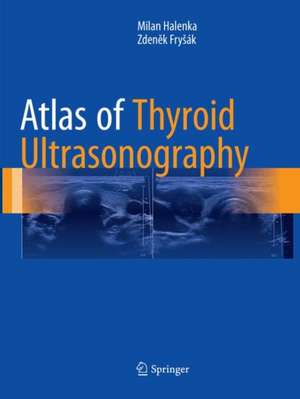
Atlas of Thyroid Ultrasonography
Autor Milan Halenka, Zdeněk Fryšáken Limba Engleză Paperback – 2 aug 2018
Including nearly 1500 ultrasound scans and covering the range of thyroid conditions, Atlas of Thyroid Ultrasonography will be a key reference for endocrinologists, radiologists, and primary care physicians, residents and fellows treating patients with thyroid problems.
| Toate formatele și edițiile | Preț | Express |
|---|---|---|
| Paperback (1) | 1864.15 lei 39-44 zile | |
| Springer International Publishing – 2 aug 2018 | 1864.15 lei 39-44 zile | |
| Hardback (1) | 2398.61 lei 39-44 zile | |
| Springer International Publishing – 16 iun 2017 | 2398.61 lei 39-44 zile |
Preț: 1864.15 lei
Preț vechi: 1962.26 lei
-5% Nou
Puncte Express: 2796
Preț estimativ în valută:
356.80€ • 371.08$ • 298.100£
356.80€ • 371.08$ • 298.100£
Carte tipărită la comandă
Livrare economică 10-15 martie
Preluare comenzi: 021 569.72.76
Specificații
ISBN-13: 9783319852379
ISBN-10: 331985237X
Pagini: 403
Ilustrații: XVIII, 403 p. 190 illus., 181 illus. in color.
Dimensiuni: 210 x 279 mm
Greutate: 11.5 kg
Ediția:Softcover reprint of the original 1st ed. 2017
Editura: Springer International Publishing
Colecția Springer
Locul publicării:Cham, Switzerland
ISBN-10: 331985237X
Pagini: 403
Ilustrații: XVIII, 403 p. 190 illus., 181 illus. in color.
Dimensiuni: 210 x 279 mm
Greutate: 11.5 kg
Ediția:Softcover reprint of the original 1st ed. 2017
Editura: Springer International Publishing
Colecția Springer
Locul publicării:Cham, Switzerland
Cuprins
Section I: Normal Thyroid Gland.- Ultrasound of Normal Thyroid Gland and Lymph Nodes.- Section II: Diffuse Thyroid Diseases .- Diffuse Goiter.- Hashimoto’s Thyroiditis: Chronic Lymphocytic Thyroiditis.- Hashimoto’s Thyroiditis: Goiter.- Hashimoto’s Thyroiditis: Atrophic Gland.- Hashimoto’s Thyroiditis: Hashitoxicosis.- Graves’ Disease.- Subacute Granulomatous Thyroiditis: de Quervain’s Diseases.- Amiodarone-induced Thyrotoxicosis.- Section III: Nodular Goiter: Benign Lesions.- Thyroid Cysts.- Solid Nodule.- Complex Nodule with Cystic Degeneration.- Complex Nodule with Calcifications.- Multinodular Goiter.- Substernal Goiter.- Toxic Multinodular Goiter and Solitary Toxic Adenoma.- Section IV: Nodular Goiter: Suspicious and Malignant Lesions.- Lesions with Intermediate Suspicion of Malignancy.- Follicular Thyroid Carcinoma.- Papillary Thyroid Microcarcinoma.- Papillary Thyroid Carcinoma: A Small Solitary Nodule ≤ 2 cm.- Papillary Thyroid Carcinoma: Medium-sized and Large Nodules > 4 cm.- Multifocal Papillary Thyroid Carcinoma.- Papillary Thyroid Carcinoma and Hashimoto’s Thyroiditis.- Papillary Thyroid Carcinoma and Graves’ Disease or Amiodarone-induced Thyrotoxicosis.- Synchronous Papillary Thyroid Carcinoma and Parathyroid Adenoma.- Differentiated Thyroid Carcinoma and Extrathyroidal Extension.- Papillary Thyroid Carcinoma in Children and Adolescents.- Medullary Thyroid Carcinoma.- Anaplastic Thyroid Carcinoma.- Other Malignancies in Thyroid Gland and Cervical Lymph Nodes: Primary Thyroid Lymphoma.- Other Malignancies in Thyroid Gland and Cervical Lymph Nodes: Extramedullary Plasmacytoma of the Thyroid Gland.- Other Malignancies in Thyroid Gland and Cervical Lymph Nodes: Malignant Cervical Lymph Nodes, Primary Outside the Thyroid Gland.- Metastatic Cervical Lymph Nodes Post Total Thyroidectomy for Thyroid Carcinoma.- Section V: Miscellanea.- Thyroid Tissue Remnants Post Thyroidectomy.- Rare Ultrasound Findings of the Thyroid Gland and Lesions Imitating Goiter.- Parathyroid Adenoma and Parathyroid Carcinoma.- Percutaneous Ethanol Injection Therapy.- Ultrasound-guided Fine-Needle Aspiration Biopsy (US-FNAB).- Thyroid Abscess as a Complication of Fine-Needle Aspiration Biopsy: A Case Report.
Recenzii
“The authors, who are from the Czech Republic, are endocrinologists and designed this atlas to be a ‘quick reference for endocrinology and radiology practices.’ It is divided into five main parts and then 25 chapters and also covers the parathyroid glands.” (Sarah Howling, RAD Magazine, February, 2018)
Notă biografică
Milan Halenka, Department of Internal Medicine III, Nephrology, Rheumatology and Endocrinology, Faculty of Medicine and Dentistry, Palacký University Olomouc, Czech Republic
Zdeněk Fryšák, Department of Internal Medicine III, Nephrology, Rheumatology and Endocrinology, Faculty of Medicine and Dentistry, Palacký University Olomouc, Czech Republic
Textul de pe ultima copertă
Combining high-quality ultrasound scans with clear and concise explanatory text, this atlas includes side-by-side depictions of various conditions of the thyroid both with and without indicative marking. Each ultrasound finding is displayed twice, six figures per page: The left-hand image is a native figure without marks; the right-hand image depicts marked findings. In this way, readers have the opportunity to see the native picture to assess it by themselves and then correct their opinion, if necessary. Five sections comprise this atlas, including the normal thyroid, diffuse thyroid lesions, both benign and malicious lesions (including various carcinomas), and rare findings.
Including nearly 1500 ultrasound scans and covering the range of thyroid conditions, Atlas of Thyroid Ultrasonography will be a key reference for endocrinologists, radiologists, and primary care physicians, residents and fellows treating patients with thyroid problems.
Including nearly 1500 ultrasound scans and covering the range of thyroid conditions, Atlas of Thyroid Ultrasonography will be a key reference for endocrinologists, radiologists, and primary care physicians, residents and fellows treating patients with thyroid problems.
Caracteristici
Combines plentiful high-quality ultrasound scans side-by-side, with markings and without, for clear depictions of indications of thyroid conditions
Covers the normal thyroid, diffuse lesions, benign and malignant lesions and goiters, and rare findings
Nearly 1500 ultrasound scans, no more than six per page
Covers the normal thyroid, diffuse lesions, benign and malignant lesions and goiters, and rare findings
Nearly 1500 ultrasound scans, no more than six per page
