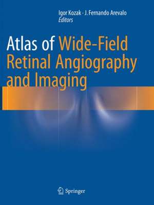
Atlas of Wide-Field Retinal Angiography and Imaging
Editat de Igor Kozak, J. Fernando Arevaloen Limba Engleză Paperback – 22 apr 2018
| Toate formatele și edițiile | Preț | Express |
|---|---|---|
| Paperback (1) | 1061.49 lei 38-44 zile | |
| Springer International Publishing – 22 apr 2018 | 1061.49 lei 38-44 zile | |
| Hardback (1) | 1626.57 lei 3-5 săpt. | |
| Springer International Publishing – 22 apr 2016 | 1626.57 lei 3-5 săpt. |
Preț: 1061.49 lei
Preț vechi: 1117.36 lei
-5% Nou
Puncte Express: 1592
Preț estimativ în valută:
203.21€ • 209.10$ • 171.29£
203.21€ • 209.10$ • 171.29£
Carte tipărită la comandă
Livrare economică 25 februarie-03 martie
Preluare comenzi: 021 569.72.76
Specificații
ISBN-13: 9783319792408
ISBN-10: 3319792407
Pagini: 259
Ilustrații: XVI, 259 p. 398 illus., 249 illus. in color.
Dimensiuni: 210 x 279 mm
Ediția:Softcover reprint of the original 1st ed. 2016
Editura: Springer International Publishing
Colecția Springer
Locul publicării:Cham, Switzerland
ISBN-10: 3319792407
Pagini: 259
Ilustrații: XVI, 259 p. 398 illus., 249 illus. in color.
Dimensiuni: 210 x 279 mm
Ediția:Softcover reprint of the original 1st ed. 2016
Editura: Springer International Publishing
Colecția Springer
Locul publicării:Cham, Switzerland
Cuprins
1. History and Principles of Wide-field Retinal Imaging.- 2. Wide-field Fluorescein Angiography.- 3. Wide-field Indocyanine Angiography.- 4. Wide-field Autofluorescence.- 5. Wide-field Retinal Imaging of Diabetic Retinopathy.- 6. Wide-field Retinal Imaging of Retinal Vascular Occlusions.- 7. Wide-field Retinal Imaging of Other Retinal Vascular Diseases.- 8. Wide-field Retinal Imaging of Retinal Dystrophies.- 9. Wide-field Retinal Imaging of Peripheral Retinal Degenerations.- 10. Wide-field Retinal Imaging of Pediatric Retina.- 11. Wide-field Retinal Imaging of Retinal and Choroidal Tumors.- 12. Wide-field Retinal Imaging of Retinal and Choroidal Inflammatory Diseases.- 13. Wide-field Retinal Imaging of Retinal and Choroidal Infectious Diseases.- 14. Wide-field Retinal Imaging of Other Miscellaneous Retinal Diseases.
Notă biografică
Igor Kozak, MD, PhD
Senior Academic Consultant
Vitreoretinal Division
King Khaled Eye Specialist Hospital
Riyadh
Saudi Arabia
J. Fernando Arevalo, MD, FACS
Edmund F. and Virginia B. Ball
Professor of Ophthalmology
Chairman, Department of Ophthalmology
Johns Hopkins Bayview Medical Center
Retina Division, Wilmer Eye Institute
The Johns Hopkins University School of Medicine
Baltimore, MD
USA
Textul de pe ultima copertă
Written by experts in the field of ophthalmology, this new text assists in understanding new imaging technology and the clinical information it can provide. It allows the reader to review numerous images of various pathologies and would be of interest to retina specialists and ophthalmologists. Retinal imaging has undergone dramatic changes in the last decade, characterized by constantly improving image resolution, most notably, as it applies to macular diseases, thanks to ever evolving optical coherence tomography. However, imaging retina outside of the vascular arcades has come a long way with advent of new cameras and lenses. The most profound change has been the introduction of wide-angle angiography, which has demonstrated pathologies previously not seen or suspected and gives rise to new theories of pathophysiology of some retinal diseases. Understanding this new imaging technology and the clinical information it can provide requires a basis of knowledge and experience in associating the findings with other clinical signs in the eye and the rest of the body. Atlas of Wide-Field Retinal Angiography and Imaging helps with this experience by allowing the reader to review numerous images of various pathologies and analyzes angiographic findings in the retinal periphery. Furthermore, as this technology increasingly fills ophthalmologists’ offices around the world, this book will prove to be an invaluable resource, written by experts in the field for retina specialists and ophthalmologists.
Caracteristici
Assists in understanding new imaging technology and the clinical information it can provide Allows the reader to review numerous images of various pathologies Analyzes angiographic findings in the retinal periphery Written by experts in the field for retina specialists and ophthalmologists Hundreds of high quality color and black and white photographs and illustrations
