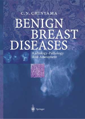
Benign Breast Diseases: Radiology — Pathology — Risk Assessment
Autor Catherine N. Chinyamaen Limba Engleză Paperback – 5 noi 2012
| Toate formatele și edițiile | Preț | Express |
|---|---|---|
| Paperback (1) | 647.01 lei 38-44 zile | |
| Springer Berlin, Heidelberg – 5 noi 2012 | 647.01 lei 38-44 zile | |
| Hardback (1) | 1175.28 lei 3-5 săpt. | |
| Springer Berlin, Heidelberg – 20 dec 2013 | 1175.28 lei 3-5 săpt. |
Preț: 647.01 lei
Preț vechi: 681.06 lei
-5% Nou
Puncte Express: 971
Preț estimativ în valută:
123.82€ • 128.48$ • 103.49£
123.82€ • 128.48$ • 103.49£
Carte tipărită la comandă
Livrare economică 13-19 martie
Preluare comenzi: 021 569.72.76
Specificații
ISBN-13: 9783642621468
ISBN-10: 3642621465
Pagini: 168
Ilustrații: XIV, 150 p.
Dimensiuni: 193 x 270 x 9 mm
Greutate: 0.39 kg
Ediția:2004
Editura: Springer Berlin, Heidelberg
Colecția Springer
Locul publicării:Berlin, Heidelberg, Germany
ISBN-10: 3642621465
Pagini: 168
Ilustrații: XIV, 150 p.
Dimensiuni: 193 x 270 x 9 mm
Greutate: 0.39 kg
Ediția:2004
Editura: Springer Berlin, Heidelberg
Colecția Springer
Locul publicării:Berlin, Heidelberg, Germany
Public țintă
Professional/practitionerCuprins
1 Radiology of Benign Breast Disease.- 2 Surgery of Benign Breast Disease.- 3 Pathology of Benign Breast Disease.- 4 Fibre-epithelial Lesions.- 5 Infiltrative Pseudo-malignant Lesions.- 6 Hyperplastic Epithelial Lesions.- 7 Cystic Lesions.- 8 Mucocele-like Lesions.- 9 Columnar Cell Lesions.- 10 Calcification in Benign Lesions.- 11 Non-epithelial Lesions.- 12 Risk Assessment in Benign Breast Disease.
Recenzii
From the reviews:
"The advent of breast screening … now means that the breast team is regularly presented with a variety of benign pathological diagnoses and an understanding of the clinical relevance of these is essential. This text aims to contribute towards this understanding. … Pathological slides are beautifully reproduced in full colour … . a comprehensive text on the pathology and risk assessment of benign breast disease which is well illustrated with pathological slides. … it would represent useful supplementary reading for non-pathologists … ." (Dr. J Litherland, RAD Magazine, July, 2005)
"Catherine Chinyama has made a creditable attempt to bring order to the often confusing field of benign breast disease. … The main focus of the book is on radiologic and pathological findings in benign breast disease … . The book is remarkably well illustrated … . The figures are informative … . The range of benign breast disease covered in the book is broad … . thisvery useful book will interest any clinician, radiologist, or pathologist who deals with benign breast disease … ." (Pamela J. Goodwin, The New England Journal of Medicine, September, 2004)
Notă biografică
Dr. Chinyama qualified with Honours Degree in Medicine in Harare, Zimbabwe, Trained in Breast Pathology at St. Bartholomew's Hospital, London and Bristol South West Breast Screening Unit in Bristol,UK. Worked as Senior Lecturer/Honorary Consultant in Histopathology at Guy's and St.Thomas' Hospital, London. Currently working as a Consultant Pathologist, Princess Elizabeth Hospital, Guernsey, Channel Islands.
Textul de pe ultima copertă
Widespread use of mammography has resulted in detection of small cancers with favorable prognosis, as well as an array of indeterminate lesions, the majority of which turn out to be histologically benign. This book gives a detailed account of the radiology and pathology of screening-detected lesions, with discussion of risk assessment, to assist clinicians in the follow-up of their patients. Related lesions are dealt with in consecutive chapters, giving the reader an opportunity for comparison. The book is well illustrated with radiological and pathological correlation of mostly screening-detected images including mucoecele-like lesions and columnar lesions. Surgeons, radiologists, pathologists and breast care nurses will find the book highly useful for the management of patients with benign or indeterminate breast lesions in a multidisciplinary setting.
Caracteristici
Includes supplementary material: sn.pub/extras
