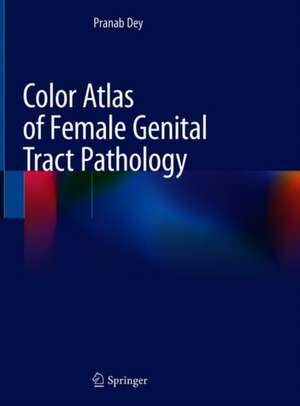
Color Atlas of Female Genital Tract Pathology
Autor Pranab Deyen Limba Engleză Hardback – 25 oct 2018
This book presents colored gross and microphotographs of histopathology sections of both common and uncommon tumors of the female genital tract, and also includes the immunohistochemistry of the important lesions. Further, it explains the salient diagnostic features and the immunocytochemistry, molecular pathology and differential diagnosis of each lesion with brief references and discusses recent advances in the diagnosis of these tumors. With numerous images offering guidance on diagnosing different lesions of the female genital tract, the book is intended for practicing pathologists and post-graduate students as well as for gynecology practitioners and post-graduate students.
| Toate formatele și edițiile | Preț | Express |
|---|---|---|
| Paperback (1) | 1238.08 lei 38-44 zile | |
| Springer Nature Singapore – 19 ian 2019 | 1238.08 lei 38-44 zile | |
| Hardback (1) | 1872.58 lei 38-44 zile | |
| Springer Nature Singapore – 25 oct 2018 | 1872.58 lei 38-44 zile |
Preț: 1872.58 lei
Preț vechi: 1971.13 lei
-5% Nou
Puncte Express: 2809
Preț estimativ în valută:
358.31€ • 375.12$ • 296.48£
358.31€ • 375.12$ • 296.48£
Carte tipărită la comandă
Livrare economică 02-08 aprilie
Preluare comenzi: 021 569.72.76
Specificații
ISBN-13: 9789811310287
ISBN-10: 9811310289
Pagini: 683
Ilustrații: XXI, 482 p. 481 illus. in color.
Dimensiuni: 210 x 279 x 32 mm
Greutate: 2.03 kg
Ediția:1st ed. 2019
Editura: Springer Nature Singapore
Colecția Springer
Locul publicării:Singapore, Singapore
ISBN-10: 9811310289
Pagini: 683
Ilustrații: XXI, 482 p. 481 illus. in color.
Dimensiuni: 210 x 279 x 32 mm
Greutate: 2.03 kg
Ediția:1st ed. 2019
Editura: Springer Nature Singapore
Colecția Springer
Locul publicării:Singapore, Singapore
Cuprins
Histopathology of vulva: inflammatory and benign neoplastic lesions.- Histopathology of vulva: pre-neoplastic and malignant tumors.- Histopathology of vagina: benign and malignant lesions.- Histopathology of cervix: benign lesions.- Histopathology of cervix: pre-neoplastic and malignant lesions.- Histopathology of endometrium: benign lesions.- Histopathology of endometrium: pre-neoplastic lesions and carcinomas.- Histopathology of uterus: mesenchymal tumors of uterus.- Histopathology of ovary: benign non neoplastic lesions.- Histopathology of ovary: epithelial tumors.- Histopathology of ovary: sex cord stromal tumors.- Histopathology of ovary: germ cell tumors.- Histopathology of ovary: metastatic and miscellaneous tumors.- Pathology of fallopian tube.- Pathology of placenta.- Histopathology of gestational trophoblastic tumors.
Notă biografică
Pranab Dey is Professor at the Department of Cytology and Gynecologic Pathology at the Post Graduate Institute of Medical Education and Research, Chandigarh. Professor Dey completed his M.D. (pathology) at the same institute and his FRCPath (cytopathology) at the Royal College of Pathologists, London. He has conducted many research projects and has pioneered work on DNA flow cytometry, image morphometry, mono-layered cytology and cytomorphologic findings of various lesions on cytology smears. He has published numerous books and articles in international journals in the field of gynecologic pathology and cytology and is a member of various societies.
Textul de pe ultima copertă
This book presents colored gross and microphotographs of histopathology sections of both common and uncommon tumors of the female genital tract, and also includes the immunohistochemistry of the important lesions. Further, it explains the salient diagnostic features and the immunocytochemistry, molecular pathology and differential diagnosis of each lesion with brief references and discusses recent advances in the diagnosis of these tumors. With numerous images offering guidance on diagnosing different lesions of the female genital tract, the book is intended for practicing pathologists and post-graduate students as well as for gynecology practitioners and post-graduate students.
Caracteristici
Includes all the common and uncommon lesions of the female genital tract
Covers the gross, histopathology, cytology, immunocytochemistry, differential diagnosis and molecular pathology of each lesion
Includes the important diagnostic features for each lesions
Includes more than 1000 images
Covers the gross, histopathology, cytology, immunocytochemistry, differential diagnosis and molecular pathology of each lesion
Includes the important diagnostic features for each lesions
Includes more than 1000 images
