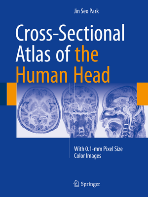
Cross-Sectional Atlas of the Human Head: With 0.1-mm pixel size color images
Autor Jin Seo Parken Limba Engleză Hardback – 19 ian 2018
| Toate formatele și edițiile | Preț | Express |
|---|---|---|
| Paperback (1) | 673.83 lei 38-44 zile | |
| Springer Nature Singapore – 7 iun 2019 | 673.83 lei 38-44 zile | |
| Hardback (1) | 1009.97 lei 38-44 zile | |
| Springer Nature Singapore – 19 ian 2018 | 1009.97 lei 38-44 zile |
Preț: 1009.97 lei
Preț vechi: 1063.13 lei
-5% Nou
Puncte Express: 1515
Preț estimativ în valută:
193.25€ • 201.79$ • 159.58£
193.25€ • 201.79$ • 159.58£
Carte tipărită la comandă
Livrare economică 11-17 aprilie
Preluare comenzi: 021 569.72.76
Specificații
ISBN-13: 9789811007699
ISBN-10: 9811007691
Pagini: 328
Ilustrații: V, 328 p. 337 illus., 313 illus. in color.
Dimensiuni: 210 x 279 mm
Greutate: 1.15 kg
Ediția:1st ed. 2017
Editura: Springer Nature Singapore
Colecția Springer
Locul publicării:Singapore, Singapore
ISBN-10: 9811007691
Pagini: 328
Ilustrații: V, 328 p. 337 illus., 313 illus. in color.
Dimensiuni: 210 x 279 mm
Greutate: 1.15 kg
Ediția:1st ed. 2017
Editura: Springer Nature Singapore
Colecția Springer
Locul publicării:Singapore, Singapore
Cuprins
Introduction.- Horizontal plane.- Coronal plane.- Sagittal plane.- Index.
Notă biografică
Jin Seo Park MD PhD. is Professor in Anatomy at Dongguk University School of Medicine.
Textul de pe ultima copertă
This superb color atlas sets a new standard in neuroanatomy by presenting around 300 detailed thin-sectioned images of the human head, including the brain, with 0.1-mm intervals and a pixel size of 0.1 mm × 0.1 mm. A new reference system employed for this purpose is clearly explained, and structures are fully annotated in the horizontal, coronal, and sagittal planes. Recent advances in 7T MRI and 7T TDI have considerably enhanced imaging of the human brain, thereby impacting on both neuroscience research and clinical practice. Moreover, the information gained from initiatives involving photography of thin slices of human cadavers, such as the Visible Human Projects, Visible Korean and Chinese Visible Human, has enriched knowledge of neuroanatomy and thereby facilitated the interpretation of such ultra-high-field resolution images. The exquisite images contained within this atlas will be invaluable in providing both researchers and clinicians with important new insights.
Caracteristici
Presents a wealth of detailed thin-sectioned color images of the human head, with a pixel size of 0.1 mm × 0.1 mm Covers all parts of the head, including the brain Provides both researchers and clinicians with important new insights


