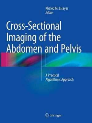
Cross-Sectional Imaging of the Abdomen and Pelvis: A Practical Algorithmic Approach
Editat de Khaled M. Elsayesen Limba Engleză Paperback – 8 oct 2016
The book is organized by anatomical organ of origin and each chapter provides a brief anatomical background of the organ under review; explores various cross-sectional imaging techniques and common pathologies; and presents practical algorithms based on frequently encountered imaging features. Special emphasis is placed on the role of computed tomography (CT) and magnetic resonance imaging (MRI). In addition to algorithmic coverage of many pathological entities in various abdominopelvic organs, unique topics are also examined, such as imaging of organ transplant (including kidney, liver and pancreas), evaluation of perianal fistula, and assessment of rectal carcinoma and prostate carcinoma by MRI. Cross-Sectional Imaging of the Abdomen and Pelvis: A Practical Algorithmic Approach is a unique and practical resource for radiologists, fellows, and residents.
| Toate formatele și edițiile | Preț | Express |
|---|---|---|
| Paperback (1) | 1284.11 lei 38-44 zile | |
| Springer – 8 oct 2016 | 1284.11 lei 38-44 zile | |
| Hardback (1) | 1898.47 lei 3-5 săpt. | |
| Springer – 27 mar 2015 | 1898.47 lei 3-5 săpt. |
Preț: 1284.11 lei
Preț vechi: 1351.70 lei
-5% Nou
Puncte Express: 1926
Preț estimativ în valută:
245.75€ • 253.87$ • 204.52£
245.75€ • 253.87$ • 204.52£
Carte tipărită la comandă
Livrare economică 22-28 martie
Preluare comenzi: 021 569.72.76
Specificații
ISBN-13: 9781493953400
ISBN-10: 1493953400
Pagini: 1066
Ilustrații: XVI, 1066 p. 1330 illus., 407 illus. in color.
Dimensiuni: 210 x 279 x 58 mm
Ediția:Softcover reprint of the original 1st ed. 2015
Editura: Springer
Colecția Springer
Locul publicării:New York, NY, United States
ISBN-10: 1493953400
Pagini: 1066
Ilustrații: XVI, 1066 p. 1330 illus., 407 illus. in color.
Dimensiuni: 210 x 279 x 58 mm
Ediția:Softcover reprint of the original 1st ed. 2015
Editura: Springer
Colecția Springer
Locul publicării:New York, NY, United States
Cuprins
Overview of CT and MRI Techniques for Evaluation of the Liver.- Diagnostic Approach of Focal and Diffuse Hepatic Diseases.- Cirrhosis and Hepatocellular Carcinoma.- Post-Locoregional Therapy Imaging of the Liver.- The Biliary Tree.- The Gall Bladder.- The Pancreas.- Cross Sectional Imaging of the Spleen.- The Stomach.- The Small Bowel.- Overview of CT Colonoscopy.- Approach to Colon Pathologies.- The Appendix.- MRI Evaluation of Rectal Carcinoma.- MRI for Perianal Fistula.- The Peritoneum.- The Extraperitoneal Spaces.- Cross Sectional Imaging of the Abdominal Wall.- Cross Sectional Imaging of the Adrenal Gland.- Prostate Imaging.- The Urinary Tract: Renal Collecting Systems, Ureters, and Urinary Bladder.- Imaging of Liver Transplant.- Imaging of Kidney Transplant.- Imaging of Pancreas Transplant.- Cross Sectional Imaging of the Kidney.- The Scrotum.- Cross Sectional Imaging of the Uterus.- The Adnexa.- The Vagina.- Cross Sectional Imaging of the Female Urethra.- Dual Energy CT and Its Applications in the Abdomen.
Notă biografică
Khaled M. Elsayes, MD, is Associate Professor of Diagnostic Radiology at the University of Texas, MD Anderson Cancer Center and University of Texas Medical School, Houston. He formerly served as Assistant Professor at the University of Michigan, Ann Arbor.
Textul de pe ultima copertă
This book offers concise descriptions of cross-sectional imaging studies of the abdomen and pelvis, supplemented with over 1100 high-quality images and discussion of state-of-the-art techniques. It is based on the most common clinical cases encountered in daily practice and uses an algorithmic approach to help radiologists arrive first at a working differential diagnosis and then reach an accurate diagnosis based on imaging features, which incorporate clinical, laboratory, and other underlying contexts.
The book is organized by anatomical organ of origin and each chapter provides a brief anatomical background of the organ under review; explores various cross-sectional imaging techniques and common pathologies; and presents practical algorithms based on frequently encountered imaging features. Special emphasis is placed on the role of computed tomography (CT) and magnetic resonance imaging (MRI). In addition to algorithmic coverage of many pathological entities in various abdominopelvic organs, unique topics are also examined, such as imaging of organ transplant (including kidney, liver and pancreas), evaluation of perianal fistula, and assessment of rectal carcinoma and prostate carcinoma by MRI. Cross-Sectional Imaging of the Abdomen and Pelvis: A Practical Algorithmic Approach is a unique and practical resource for radiologists, fellows, and residents.
The book is organized by anatomical organ of origin and each chapter provides a brief anatomical background of the organ under review; explores various cross-sectional imaging techniques and common pathologies; and presents practical algorithms based on frequently encountered imaging features. Special emphasis is placed on the role of computed tomography (CT) and magnetic resonance imaging (MRI). In addition to algorithmic coverage of many pathological entities in various abdominopelvic organs, unique topics are also examined, such as imaging of organ transplant (including kidney, liver and pancreas), evaluation of perianal fistula, and assessment of rectal carcinoma and prostate carcinoma by MRI. Cross-Sectional Imaging of the Abdomen and Pelvis: A Practical Algorithmic Approach is a unique and practical resource for radiologists, fellows, and residents.
Caracteristici
CT and MR protocols are described for each abdominal organ Clinical cases are accompanied by imaging features and illustrated algorithms to reinforce pattern recognition for interpretation and diagnosis Contains state-of-the art technique descriptions
