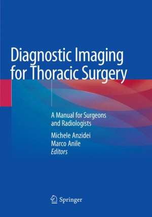
Diagnostic Imaging for Thoracic Surgery: A Manual for Surgeons and Radiologists
Editat de Michele Anzidei, Marco Anileen Limba Engleză Paperback – 16 feb 2019
| Toate formatele și edițiile | Preț | Express |
|---|---|---|
| Paperback (1) | 728.16 lei 22-36 zile | |
| Springer International Publishing – 16 feb 2019 | 728.16 lei 22-36 zile | |
| Hardback (1) | 889.51 lei 38-44 zile | |
| Springer International Publishing – 3 sep 2018 | 889.51 lei 38-44 zile |
Preț: 728.16 lei
Preț vechi: 766.49 lei
-5% Nou
Puncte Express: 1092
Preț estimativ în valută:
139.38€ • 151.45$ • 117.15£
139.38€ • 151.45$ • 117.15£
Carte disponibilă
Livrare economică 31 martie-14 aprilie
Preluare comenzi: 021 569.72.76
Specificații
ISBN-13: 9783030078881
ISBN-10: 3030078884
Pagini: 358
Ilustrații: VIII, 358 p. 350 illus., 137 illus. in color.
Dimensiuni: 178 x 254 mm
Greutate: 0.73 kg
Ediția:Softcover reprint of the original 1st ed. 2018
Editura: Springer International Publishing
Colecția Springer
Locul publicării:Cham, Switzerland
ISBN-10: 3030078884
Pagini: 358
Ilustrații: VIII, 358 p. 350 illus., 137 illus. in color.
Dimensiuni: 178 x 254 mm
Greutate: 0.73 kg
Ediția:Softcover reprint of the original 1st ed. 2018
Editura: Springer International Publishing
Colecția Springer
Locul publicării:Cham, Switzerland
Cuprins
Pre-operative and post-operative chest X-Ray.- Indications to The Use of Computed Tomography in Thoracic Pathologies.- PET Hybrid Imaging of the Thorax.- Chest MRI.- Interventional radiology procedures.- Normal radiologic anatomy and anatomical variants of the chest relevant to thoracic surgery.- Identification and characterization of lung nodules.- Staging of non-small-cell lung cancer.- Staging of Small Cell Lung Cancer.- Staging of Mesothelioma.- Imaging of non-neoplastic lung diseases requiring a surgical management.- Lung and Airways surgical procedures.- Imaging and staging of thymic tumors.- Imaging of Nonthymic Anterior Mediastinal Masses.- Imaging of non-neoplastic mediastinal pathologies.- Imaging and staging of esophageal tumors.- Imaging of non-neoplastic esophageal pathologies.- Imaging of chest wall tumors.- Imaging of non-neoplastic chest wall pathologies.- Imaging of post-surgical complications in the lung and mediastinum.
Recenzii
“This is an up-to-date, complete reference on multimodality imaging of the nonvascular thorax. The information is presented in a logical and efficient format. The authors are successful in creating a practical book that is useful for all disciplines involved in the diagnosis and management of thoracic disease. … This is useful for residents, fellows, and practitioners. The book targets medical and surgical practitioners, but, as a radiology resident, I have found it very useful.” (Robert Harrold, Doody's Book Reviews, January 04, 2019)
Notă biografică
Michele Anzidei is a body radiologist at the Department of Radiological, Oncological, and Anatomopathological Sciences of the Sapienza University in Rome, Italy. Dr. Anzidei completed a PhD in cardio-angio-thoracic imaging and was a research fellow in oncologic molecular imaging for 2 years prior to being appointed Assistant Professor. He has a particular interest in chest CT and lung interventions. He is a member of the European Society of Radiology (ESR) and has been an invited lecturer at several national and international congresses. Dr. Anzidei is an editorial board member or reviewer for various scientific journals and has authored more than 60 scientific papers and several book chapters.
Marco Anile is a thoracic surgeon surgeon at the Department of Surgery and Organ Transplantation of the Sapienza University in Rome, Italy. Dr. Anile completed a PhD in cardio-angio-thoracic physiopathology and was appointed Assistant Professorin 2009; he is currently Associate Professor of Thoracic Surgery. He has a particular interest in chest oncology, respiratory insufficiency, and lung transplantation. He is a member of European Society of Thoracic Surgeons and has been an invited lecturer at a number of national and international congresses. Dr. Anile is an editorial board member or reviewer for various scientific journals. He has authored more than 100 scientific papers and several book chapters.
Marco Anile is a thoracic surgeon surgeon at the Department of Surgery and Organ Transplantation of the Sapienza University in Rome, Italy. Dr. Anile completed a PhD in cardio-angio-thoracic physiopathology and was appointed Assistant Professorin 2009; he is currently Associate Professor of Thoracic Surgery. He has a particular interest in chest oncology, respiratory insufficiency, and lung transplantation. He is a member of European Society of Thoracic Surgeons and has been an invited lecturer at a number of national and international congresses. Dr. Anile is an editorial board member or reviewer for various scientific journals. He has authored more than 100 scientific papers and several book chapters.
Textul de pe ultima copertă
This book offers a comprehensive overview of thoracic pathologies of surgical interest involving the lung, mediastinum, esophagus, and chest wall with the aim of providing both radiologists and thoracic surgeons with a reference of high value in everyday clinical practice. Oncologic and non-oncologic conditions are reviewed from both the radiological and the surgical point of view, each one being documented with the aid of high-quality radiologic images from several modalities (including X-ray, fluoroscopy, CT, MR, and PET), illustrations/artwork, and high-definition images from the surgical table. The postoperative anatomy and complications associated with thoracic surgery procedures are also described in detail, with provision of imaging examples that highlight aspects of importance in differentiating between normal and abnormal findings. Written by experts in the field, Diagnostic Imaging for Thoracic Surgery is exceptional in combining precise descriptions of surgical procedures with key teaching points in imaging interpretation.
Caracteristici
Describes in detail imaging and surgical procedures in lung, mediastinal, and chest wall pathologies Aids in differentiating between normal postoperative anatomy and complications Features numerous high-quality radiologic images and high-definition images from the surgical table
