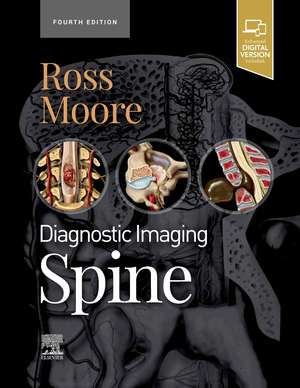
Diagnostic Imaging: Spine: Diagnostic Imaging
Autor Jeffrey S. Ross, Kevin R. Mooreen Limba Engleză Hardback – 13 dec 2020
Din seria Diagnostic Imaging
- 5%
 Preț: 1654.43 lei
 - 5%
 Preț: 1618.70 lei
 - 25%
 Preț: 1644.20 lei
 - 24%
 Preț: 1710.90 lei
 - 5%
 Preț: 906.63 lei
 - 25%
 Preț: 1644.12 lei
 - 5%
 Preț: 1435.85 lei
 - 24%
 Preț: 1676.28 lei
 - 5%
 Preț: 731.07 lei
 - 5%
 Preț: 1306.73 lei
 - 5%
 Preț: 349.24 lei
 - 5%
 Preț: 1462.37 lei
 - 5%
 Preț: 1108.87 lei
 - 24%
 Preț: 1649.37 lei
 - 24%
 Preț: 1711.38 lei
 - 5%
 Preț: 720.68 lei
 - 24%
 Preț: 1679.91 lei
 - 24%
 Preț: 1671.88 lei
 - 24%
 Preț: 1670.08 lei
 - 5%
 Preț: 1307.85 lei
 - 5%
 Preț: 1308.02 lei
 - 5%
 Preț: 383.93 lei
 - 5%
 Preț: 663.23 lei
 - 5%
 Preț: 1437.67 lei
 - 5%
 Preț: 1130.07 lei
 - 5%
 Preț: 717.20 lei
 - 5%
 Preț: 1953.34 lei
 - 5%
 Preț: 1450.84 lei
 - 5%
 Preț: 1626.03 lei
 - 5%
 Preț: 794.00 lei
 - 24%
 Preț: 1664.93 lei
 - 5%
 Preț: 1113.99 lei
 - 5%
 Preț: 1184.42 lei
 - 5%
 Preț: 1308.74 lei
 - 24%
 Preț: 1669.16 lei
 - 5%
 Preț: 1101.21 lei
 - 5%
 Preț: 821.19 lei
 - 24%
 Preț: 1667.83 lei
 - 5%
 Preț: 1950.60 lei
 - 5%
 Preț: 783.04 lei
 - 5%
 Preț: 975.17 lei
 - 5%
 Preț: 1105.61 lei
 - 5%
 Preț: 1301.44 lei
 - 5%
 Preț: 1116.00 lei
 - 5%
 Preț: 1605.11 lei
 - 5%
 Preț: 802.21 lei
 - 5%
 Preț: 1113.11 lei
 - 5%
 Preț: 1298.14 lei
Preț: 1653.20 lei
Preț vechi: 2185.41 lei
-24% Nou
316.38€ • 329.09$ • 261.19£
Carte disponibilă
Livrare economică 17-31 martie
Specificații
ISBN-10: 0323793991
Pagini: 1256
Dimensiuni: 216 x 276 x 57 mm
Greutate: 2.68 kg
Ediția:4
Editura: Elsevier
Seria Diagnostic Imaging
Cuprins
Congenital and Genetic Disorders
Congenital
1 Normal Anatomical Variations
2 Normal Anatomy
3 Measurement Techniques
4 MR Artifacts
5 Normal Variant
6 Craniovertebral Junction Variants
7 Ponticulus Posticus
8 Ossiculum Terminale
9 Conjoined Nerve Roots
10 Limbus Vertebra
11 Filum Terminale Fibrolipoma
12 Bone Island
13 Ventriculus Terminalis
Chiari Disorders
14 Chiari 0
15 Chiari 1
16 Complex Chiari
17 Chiari 2
18 Chiari 3
Abnormalities of Neurulation
19 Approach to Spine and Spinal Cord Development
20 Myelomeningocele
21 Lipomyelomeningocele
22 Lipoma
23 Dorsal Dermal Sinus
24 Simple Coccygeal Dimple
25 Dermoid Cysts
26 Epidermoid Cysts
Anomalies of the Caudal Cell Mess
27 Tethered Spinal Cord
28 Segmental Spinal Dysgenesis
29 Caudal Regression Syndrome
30 Terminal Myelocystocele
31 Anterior Sacral Meningocele
32 Sacral Extradural Arachnoid Cyst
33 Sacrococcygeal Teratoma
Anomalies of Notochord and Vertebral Formation
34 Craniovertebral Junction Embryology
35 Paracondylar Process
36 Split Atlas
37 Klippel-Feil Spectrum
38 Failure of Vertebral Formation
39 Vertebral Segmentation Failure
40 Split Cord Malformation (formerly diastematomyelia)
41 Partial Vertebral Duplication
42 Incomplete Fusion, Posterior Element
43 Neurenteric Cyst
Developmental Abnormalities
44 Os Odontoideum
45 Lateral Meningocele
46 Dorsal Spinal Meningocele
47 Dural Dysplasia
48 Genetic Disorders
49 Neurofibromatosis Type 1
50 Neurofibromatosis Type 2
51 Schwannomatosis
52 Achondroplasia
53 Mucopolysaccharidoses
54 Sickle Cell Disease
55 Osteogenesis Imperfecta
56 Tuberous Sclerosis
57 Osteopetrosis
58 Gaucher Disease
59 Ochronosis
60 Connective Tissue Disorders
61 Spondyloepiphyseal Dysplasia
DEL Thanatophoric Dwarfism
Scoliosis and Kyphosis
62 Introduction to Scoliosis
63 Scoliosis
64 Kyphosis
65 Degenerative Scoliosis
66 Flat Back Syndrome
67 Scoliosis Instrumentation
Trauma
68 Vertebral Column, Discs, and Paraspinal Muscle
69 Fracture Classification
70 Atlantooccipital Dislocation
71 Ligamentous Injury
72 Occipital Condyle Fracture
73 Jefferson C1 Fracture
74 Atlantoaxial Rotatory Fixation
75 Odontoid C2 Fracture
76 Burst C2 Fracture
77 Hangman's C2 Fracture
78 Apophyseal Ring Fracture
79 Cervical Hyperflexion Injury
80 Cervical Hyperextension Injury
81 Cervical Hyperextension-Rotation Injury
82 Cervical Burst Fracture
83 Cervical Hyperflexion-Rotation Injury
84 Cervical Lateral Flexion Injury
85 Cervical Posterior Column Injury
86 Traumatic Disc Herniation
87 Thoracic and Lumbar Burst Fracture
88 Facet-Lamina Thoracolumbar Fracture
89 Fracture Dislocation
90 Chance Fracture
91 Thoracic and Lumbar Hyperextension Injury
92 Anterior Compression Fracture
93 Lateral Compression Fracture
94 Lumbar Facet-Posterior Fracture
95 Sacral Traumatic Fracture
96 Pedicle Stress Fracture
97 Sacral Insufficiency Fracture
Cord, Dura, and Vessels
98 SCIWORA
99 Post-Traumatic Syrinx
100 Presyrinx Edema
101 Spinal Cord Contusion-Hematoma
102 Idiopathic Spinal Cord Herniation
103 Central Spinal Cord Syndrome
104 Traumatic Dural Tear
105 Traumatic Epidural Hematoma
106 Traumatic Subdural Hematoma
107 Vascular Injury, Cervical
108 Traumatic Arteriovenous Fistula
109 Wallerian Degeneration
Degenerative Diseases and Arthritides
Degenerative Diseases
110 Nomenclature of Degenerative Disc Disease
111 Degenerative Disc Disease
112 Degenerative Endplate Changes
113 Degenerative Arthritis of the CVJ
114 Disc Bulge
115 Anular Fissure, Intervertebral Disc
116 Cervical Intervertebral Disc Herniation
117 Thoracic Intervertebral Disc Herniation
118 Lumbar Intervertebral Disc Herniation
119 Intervertebral Disc Extrusion, Foraminal
120 Cervical Facet Arthropathy
121 Lumbar Facet Arthropathy
122 Facet Joint Synovial Cyst
123 Baastrup Disease
124 Bertolotti Syndrome
125 Schmorl Node
126 Scheuermann Disease
127 Acquired Lumbar Central Stenosis
128 Congenital Spinal Stenosis
129 Cervical Spondylosis
130 DISH
131 OPLL
132 Ossification Ligamentum Flavum
133 Periodontoid Pseudotumor
Spondylolisthesis and Spondylolysis
134 Spondylolisthesis
135 Spondylolysis
136 Instability
Inflammatory, Crystalline, and Miscellaneous Arthritides
137 Adult Rheumatoid Arthritis
138 Juvenile Idiopathic Arthritis
139 Spondyloarthropathy
140 Neurogenic (Charcot) Arthropathy
141 Hemodialysis Spondyloarthropathy
142 Ankylosing Spondylitis
143 CPPD
144 Gout
145 Longus Colli Calcific Tendinitis
Infection and Inflammatory Disorders
Infections
146 Pathways of Spread
147 Spinal Meningitis
148 Pyogenic Osteomyelitis
149 Tuberculous Osteomyelitis
150 Fungal and Miscellaneous Osteomyelitis
151 Osteomyelitis, C1-C2
152 Brucellar Spondylitis
153 Septic Facet Joint Arthritis
154 Paraspinal Abscess
155 Epidural Abscess
156 Subdural Abscess
157 Abscess, Spinal Cord
158 Viral Myelitis
159 HIV Myelitis
160 Syphilitic Myelitis
161 Opportunistic Infections
162 Echinococcosis
163 Schistosomiasis
164 Cysticercosis
Inflammatory and Autoimmune Disorders
165 Acute Transverse Myelopathy
166 Idiopathic Acute Transverse Myelitis
167 Multiple Sclerosis
168 Neuromyelitis Optica SPECTRUM DISORDER
169 ADEM
170 Guillain-Barré Syndrome
171 CIDP
172 Chronic Recurrent Multifocal Osteomyelitis
173 Grisel Syndrome
174 Paraneoplastic Myelopathy
175 IgG4-Related Disease/Hypertrophic Pachymeningitis
Neoplasms, Cysts, and Other Masses
Neoplasms
176 Introduction and Overview
177 Spread of Neoplasms
178 SINS, NOMS and ESCC
Extradural
179 Imaging of metastatic disease prose intro
180 Blastic Osseous Metastases
181 Lytic Osseous Metastases
182 Hemangioma
183 Osteoid Osteoma
184 Osteoblastoma
185 Aneurysmal Bone Cyst
186 Giant Cell Tumor
187 Osteochondroma
188 Chondrosarcoma
189 Osteosarcoma
190 Chordoma
191 Ewing Sarcoma
192 Lymphoma
193 Leukemia
194 Plasmacytoma
195 Multiple Myeloma
196 Neuroblastic Tumor
197 Langerhans Cell Histiocytosis
198 Angiolipoma
Intradural Extramedullary
199 Schwannoma
200 Melanotic Schwannoma
201 Meningioma
202 Solitary Fibrous Tumor/Hemangiopericytoma
203 Neurofibroma
204 Malignant Nerve Sheath Tumors
205 Metastases, CSF Disseminated
206 Paraganglioma
207 Intramedullary
208 Astrocytoma
209 Cellular Ependymoma
210 Myxopapillary Ependymoma
211 Hemangioblastoma
212 Spinal Cord Metastases
213 Primary Melanocytic Neoplasms/Melanocytoma
214 Ganglioglioma
Nonneoplastic Cysts, Tumor Mimics and CSF Disorders
Cysts
215 CSF Flow Artifact
216 Meningeal Cyst
217 Perineural Root Sleeve Cyst
218 Syringomyelia
Nonneoplastic Masses and Tumor Mimics
219 Epidural Lipomatosis
220 Normal Fatty Marrow Variants
221 Fibrous Dysplasia
222 CAPNON
223 Kümmell Disease
224 Hirayama Disease
225 CSF leak disorders
226 SIH
227 Ventral defects / fast leak
228 Root sleeve leaks
229 CSF venous fistula
Vascular
Vascular Anatomy and Congenital Lesions
230 Vascular Anatomy
231 Persistent First Intersegmental Artery
232 Persistent Hypoglossal Artery
234 Persistent Proatlantal Artery
Vascular Malformations
235 Type 1 Vascular Malformation (dAVF)
236 Type 2 Arteriovenous Malformation (AVM)
237 Type 3 Arteriovenous Malformation (AVM)
238 Type 4 Vascular Malformation (AVF)
239 Conus Arteriovenous Malformation
240 Posterior Fossa Dural Fistula With Intraspinal Drainage
241 Epidural fistula
242 Cavernous Malformation
Vascular Misc
243 Spinal Artery Aneurysm
244 Spinal Cord Infarction
245 Subarachnoid Hemorrhage
246 Spontaneous Epidural Hematoma
247 Subdural Hematoma
248 Superficial Siderosis
249 Hematomyelia/Nontraumatic Cord Hemorrhage
250 Bow Hunter Syndrome
251 Vertebral Artery Dissection
Systemic Disorders
252 Spinal Manifestations of Systemic Diseases
253 Osteoporosis
254 Paget Disease
255 Hyperparathyroidism
256 Renal Osteodystrophy
257 Hyperplastic Vertebral Marrow
258 Myelofibrosis
259 Bone Infarction
260 Extramedullary Hematopoiesis
261 Tumoral Calcinosis
262 Sarcoidosis
263 Subacute Combined Degeneration
Peripheral Nerve and Plexus
Plexus and Peripheral Nerve Lesions
264 Normal Plexus and Nerve Anatomy
265 Superior Sulcus Tumor
266 Thoracic Outlet Syndrome
267 Muscle Denervation
268 Brachial Plexus Traction Injury
269 Idiopathic Brachial Plexus Neuritis
270 Traumatic Neuroma
271 Radiation Plexopathy
272 Peripheral Nerve Sheath Tumor
273 Peripheral Neurolymphomatosis
274 Hypertrophic Neuropathy
275 Femoral Neuropathy
276 Ulnar Neuropathy
277 Suprascapular Neuropathy
278 Median Neuropathy
279 Common Peroneal Neuropathy
280 Tibial Neuropathy
Spine Postprocedural Imaging
281 Postoperative Imaging and Complications
282 Surgical Approaches
283 Normal Postoperative Change
284 Postoperative Spinal Complications
285 Myelography Complications
286 Vertebroplasty Complications
287 Failed Back Surgery Syndrome
288 Recurrent Disc Herniation
289 Peridural Fibrosis
290 Arachnoiditis/Adhesions
291 Arachnoiditis Ossificans
292 Accelerated Degeneration
293 Postoperative Infection
294 Pseudomeningocele
295 CSF Leakage Syndrome
296 Postsurgical Deformity
Hardware
297 Metal Artifact
298 Occipitocervical Fixation
299 Plates and Screws
300 Cages
301 Interbody Fusion Devices
302 Interspinous Spacing Devices
303 Cervical Artificial Disc
304 Lumbar Artificial Disc
305 Hardware Failure
306 Bone Graft Complications
307 rhBMP-2 Complications
308 Heterotopic Bone Formation
Post Radiation and Chemotherapy Complications
309 Radiation Myelopathy
310 Post-Irradiation Vertebral Marrow
311 Anterior Lumbar Radiculopathy
Descriere
Covering the entire spectrum of this fast-changing field, Diagnostic Imaging: Spine, fourth edition, is an invaluable resource for general radiologists, neuroradiologists, and trainees-anyone who requires an easily accessible, highly visual reference on today's spinal imaging. Drs. Jeffrey Ross, Kevin Moore, and their team of highly regarded experts provide updated information on disease identification and imaging techniques to help you make informed decisions at the point of care. The text is lavishly illustrated, delineated, and referenced, making it a useful learning tool as well as a handy reference for daily practice.
