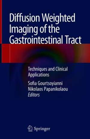
Diffusion Weighted Imaging of the Gastrointestinal Tract: Techniques and Clinical Applications
Editat de Sofia Gourtsoyianni, Nikolaos Papanikolaouen Limba Engleză Hardback – 26 oct 2018
The book reviews the technical aspects and clinical applications of DWI in imaging of the GI tract, and provides specific technical details (imaging protocols, artefacts, optimization techniques) for each GI tract division. This volume is mainly intended for radiologists who are interested in abdominal radiology, as well as radiology residents. Given that magnetic resonance physics is complex and can be cumbersome to learn, the authors have made it as simple and practical as possible.
| Toate formatele și edițiile | Preț | Express |
|---|---|---|
| Paperback (1) | 321.62 lei 38-44 zile | |
| Springer International Publishing – 13 dec 2018 | 321.62 lei 38-44 zile | |
| Hardback (1) | 335.17 lei 38-44 zile | |
| Springer International Publishing – 26 oct 2018 | 335.17 lei 38-44 zile |
Preț: 335.17 lei
Preț vechi: 352.81 lei
-5% Nou
Puncte Express: 503
Preț estimativ în valută:
64.15€ • 69.71$ • 53.93£
64.15€ • 69.71$ • 53.93£
Carte tipărită la comandă
Livrare economică 16-22 aprilie
Preluare comenzi: 021 569.72.76
Specificații
ISBN-13: 9783319928180
ISBN-10: 331992818X
Pagini: 97
Ilustrații: VII, 85 p. 38 illus., 8 illus. in color.
Dimensiuni: 155 x 235 mm
Greutate: 0.32 kg
Ediția:1st ed. 2019
Editura: Springer International Publishing
Colecția Springer
Locul publicării:Cham, Switzerland
ISBN-10: 331992818X
Pagini: 97
Ilustrații: VII, 85 p. 38 illus., 8 illus. in color.
Dimensiuni: 155 x 235 mm
Greutate: 0.32 kg
Ediția:1st ed. 2019
Editura: Springer International Publishing
Colecția Springer
Locul publicării:Cham, Switzerland
Cuprins
DWI sequences for GI tract Imaging .- Upper GI Tract (oesophagus/stomach/duodemum) – Simon Jackson.- Small bowel - IBD (Crohn's disease).- Small bowel - Tumours (e.g. carcinoid, lymphoma,serosal disease from other primaries)- Phillippe Soyer/ Christine Hoeffel.- Large bowel (Diverticulitis vs sigmoid cancer, polyps , MRColonography?).- Rectum (rectal cancer- pre/post..)- Regina Beets Tan and coworkers.- Anal canal (anal cancer)- Sofia Gourtsoyianni and Vicky Goh.
Notă biografică
Sofia Gourtsoyianni obtained her Medical Degree from the Medical School of the University of Pecs, Hungary in 2001. She spent the first 18 months of her Radiology residency at the Institute of Clinical Radiology, Ludwig Maximilian University, Munich, Germany, and then continued at the Radiology Department of the University Hospital of Heraklion, Greece.
In June 2010 she obtained her PhD from the University of Heraklion, Crete, Greece on Diffusion Weighted Magnetic Resonance Imaging in Staging of Rectal Cancer.
She completed a one-year Research Fellowship in CT Applications at the Department of Computed Tomography, Beth Israel Deaconess Medical Center, Harvard Medical School, Boston, USA in 2011.
During the period 2013-2017 she worked as a Consultant in Radiology at Guy’s & St Thomas’ NHS Foundation Trust after having spent a year as a Clinical Research Fellow at the Division of Cancer Imaging, School of Biomedical Engineering and Imaging Sciences (BMEIS), King’s College London working with Prof Vicky Goh. She currently holds an Honorary Clinical Lecturer appointment with King’s College London and works as a Consultant Radiologist at Konstantopouleion General Hospital in Athens, Greece.
Dr. Gourtsoyianni’s main areas of clinical and research interest are abdominal/GI and oncologic imaging. She has (co) authored 28 peer reviewed papers and 7 book chapters. She has delivered more than 80 national/international invited lectures at radiological and clinical society meetings around the world.
Nikolaos Papanikolaou is the Head of Computational Clinical Imaging Group at Champalimaud Foundation in Lisbon, and an affiliated researcher of the Karolinska Institute in Stockholm. He received his Ph.D. in Medicine from the University of Crete, Greece. He has been actively involved on clinical MRI research for the last 25 years.
Dr. Papanikolaou has published 63 papers in international peer reviewed journals, authored 18 book chapters, and held 81 invited lectures for international congresses and educational courses. He is an honorary member of SEDIA and a fellow member of ESGAR. His main research focus is on Diffusion Weighted Imaging, Radiomics, Modelling and Post Processing techniques.
Textul de pe ultima copertă
This book explains how diffusion weighted imaging has been incorporated in routine MRI examinations of the abdomen and pelvis: though its clinical role is still evolving, it is already considered an important tool for the assessment of rectal cancer treatment response, as was confirmed in recent ESGAR consensus statements. The standardization and clinical validation of quantitative DWI related biomarkers are still in progress, although certain efforts have been undertaken to establish imaging guidelines for different clinical indications/body parts.
The book reviews the technical aspects and clinical applications of DWI in imaging of the GI tract, and provides specific technical details (imaging protocols, artefacts, optimization techniques) for each GI tract division. This volume is mainly intended for radiologists who are interested in abdominal radiology, as well as radiology residents. Given that magnetic resonance physics is complex and can be cumbersome to learn, the
authors have made it as simple and practical as possible.
The book offers a useful tool for radiologists with a particular interest in the gastrointestinal tract radiology, as well as for radiology residents.
authors have made it as simple and practical as possible.
The book offers a useful tool for radiologists with a particular interest in the gastrointestinal tract radiology, as well as for radiology residents.
Caracteristici
Discusses both the technical aspects and clinical applications of Diffusion Weighted Imaging Presents an overview of the core principles and technical aspects of Diffusion Weighted Imaging in the GI tract Highlights pearls and pitfalls concerning Diffusion Weighted Imaging in the GI tract
