Cantitate/Preț
Produs

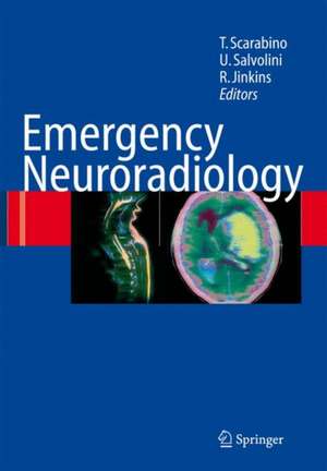
Emergency Neuroradiology

Editat de Tommaso Scarabino, Ugo Salvolini, Randy J. Jinkins
en Limba Engleză Hardback – 22 noi 2005
Encouraged by the success of the Italian editions, the Authors have decided to publish an English version taking into account the latest technical and methodological advances and the consequent new acquisitions in clinical practice. The contribution of Professor R. Jinkins has been essential to carry out both these tasks. The resulting work is an up-to-date technical tool that preserves its original aim of contributing to the training of those radiologists who work in emergency departments. We hope that this revised and extended English version will have the same success as the pre- ous Italian editions, thereby confirming the validity of our initiative. The work of all the friends and colleagues who have contributed to the making of this book is gratefully acknowledged. Tommaso Scarabino Ugo Salvolini CONTENTS I. CEREBROVASCULAR EMERGENCIES 1. 1 Clinical and diagnostic summary Neuroradiological protocol . . . . . . . . . . . . . . . . . . . . . . . . . . . . . . . . . . . . . . . . . . . . . . . . . . . . . . . . . . . . . . . . . . . 3 Haemorrhagic stroke . . . . . . . . . . . . . . . . . . . . . . . . . . . . . . . . . . . . . . . . . . . . . . . . . . . . . . . . . . . . . . . . . . . . . . . . . . 4 Ischaemic stroke . . . . . . . . . . . . . . . . . . . . . . . . . . . . . . . . . . . . . . . . . . . . . . . . . . . . . . . . . . . . . . . . . . . . . . . . . . . . . . . . 4 Conclusions . . . . . . . . . . . . . . . . . . . . . . . . . . . . . . . . . . . . . . . . . . . . . . . . . . . . . . . . . . . . . . . . . . . . . . . . . . . . . . . . . . . . . 6 References . . . . . . . . . . . . . . . . . . . . . . . . . . . . . . . . . . . . . . . . . . . . . . . . . . . . . . . . . . . . . . . . . . . . . . . . . . . . . . . . . . . . . . . 6 1. 2 CT in ischaemia Introduction . . . . . . . . . . . . . . . . . . . . . . . . . . . . . . . . . . . . . . . . . . . . . . . . . . . . . . . . . . . . . . . . . . . . . . . . . . . . . . . . . . . . 7 Semeiotics . . . . . . . . . . . . . . . . . . . . . . . . . . . . . . . . . . . . . . . . . . . . . . . . . . . . . . . . . . . . . . . . . . . . . . . . . . . . . . . . . . . . . . . 7 Particular forms of infarction . . . . . . . . . . . . . . . . . . . . . . . . . . . . . . . . . . . . . . . . . . . . . . . . . . . . . . . . . . . . . . . . . 21 Differential diagnosis . . . . . . . . . . . . . . . . . . . . . . . . . . . . . . . . . . . . . . . . . . . . . . . . . . . . . . . . . . . . . . . . . . . . . . . . . . 24 Conclusions . . . . . . . . . . . . . . . . . . . . . . . . . . . . . . . . . . . . . . . . . . . . . . . . . . . . . . . . . . . . . . . . . . . . . . . . . . . . . . . . . . . . . 25 References . . . . . . . . . . . . . . . . . . . . . . . . . . . . . . . . . . . . . . . . . . . . . . . . . . . . . . . . . . . . . . . . . . . . . . . . . . . . . . . . . . . . . . . 25 1. 3 CT in intraparenchymal haemorrhage Introduction . . . . . . . . . . . . . . . . . . . . . . . . . . . . . . . . . . . . . . . . . . . . . . . . . . . . . . . . . . . . . . . . . . . . . . . . . . . . . . . . . . . . 27 The role of CT . . . . . . . . . . . . . . . . . . . . . . . . . . . . . . . . . . . . . . . . . . . . . . . . . . . . . . . . . . . . . . . . . . . . . . . . . . . . . . . . . . 28 Causes . . . . . . . . . . . . . . . . . . . . . . . . . . . . . . . . . . . . . . . . . . . . . . . . . . . . . . . . . . . . . . . . . . . . . . . . . . . . . . . . . . . . . . . . . . . 29 Semeiotics . . . . . . . . . . . . . . . . . . . . . . . . . . . . . . . . . . . . . . . . . . . . . . . . . . . . . . . . . .. . . . . . . . . . . . . . . . . . . . . . . . . . . . . 34 Particular forms of IPH . . . . . . . . . . . . . . . . . . . . . . . . . . . . . . . . . . . . . . . . . . . . . . . . . . . . . . . . . . . . . . . . . . . . . . . 46 Conclusions . . . . . . . . . . . . . . . . . . . . . . . . . . . . . . . . . . . . . . . . . . . . . . . . . . . . . . . . . . . . . . . . . . . . . . . . . . . . . . . . . . . . . 47 References . . . . . . . . . . . . . . . . . . . . . . . . . . . . . . . . . . . . . . . . . . . . . . . . . . . . . . . . . . . . . . . . . . . . . . . . . . . . . . . . . . . . . . . 47 1. 4 CT use in subarachnoid haemorrhage Introduction . . . . . . . . . . . . . . . . . . . . . . . . . . . . . . . . . . . . . . . . . . . . . . . . . . . . . . . . . . . . . . . . . . . . . . . . . . . . . . . . . . . . 49 Semeiotics . . . . . . . . . . . . . . . . . . . . . . . . . . . . . . . . . . . . . . . . . . . . . . . . . . . . . . . . . . . . . . . . . . . . . . . . . . . . . . . . . . . . . . . 49 Causes . . . . . . . . . . . . . . . . . . . . . . . . . . . . . . . . . . . . . . . . . . . . . . . . . . . . . . . . . . . . . . . . . . . . . . . . . . . . . . . . . . . . . . . . . . . 52 Complications . . . . . . . . . . . . . . . . . . . . . . . . . . . . . . . . . . . . . . . . . . . . . . . . . . . . . . . . . . . . . . . . . . . . . . . . . . . . . . . . . . 58 Postsurgical follow-up . . . . . . . . . . . . . . . . . . . . . . . . . . . . . . . . . . . . . . . . . . . . . . . . . . . . . . . . . . . . . . . . . . . . . . . . . 59 Conclusions . . . . . . . . . . . . . . . . . . . . . . . . . . . . . . . . . . . . . . . . . . . . . . . . . . . . . . . . . . . . . . . . . . . . . . . . . . . . . . . . . . . . . 60 References . . . . . . . . . . . . . . . . . . . . . .. . . . . . . . . . . . . . . . . . . . . . . . . . . . . . . . . . . . . . . . . . . . . . . . . . . . . . . . . . . . . . . . . 60 1. 5 MRI in ischaemia Introduction . . . . . . . . . . . . . . . . . . . . . . . . . . . . . . . . . . . . . . . . . . . . . . . . . . . . . . . . . . . . . . . . . . . . . . . . . . . . . . . . . . . . 63 Semeiotics . . . . . . . . . . . . . . . . . . . . . . . . . . . . . . . . . . . . . . . . . . . . . . . . . . . . . . . . . . . . . . . . . . . . . . . . . . . . . . . . . . . . . . . 64 Possible uses of clinical MR in the diagnosis of emergency ischaemia after the hyperacute phase . . . . . . . . . . . . . . . . . . . . . . . . . . . . . . . . . . . . . . . . . . . . . . . . . . . . . . . . . . . . . . . . . . . 70 References . . . . . . . . . . . . . . . . . . . . . . . . . . . . . . . . . . . . . . . . . . . . . . . . . . . . . . . . . . . . . . . . . . . . . . . . . . . . . . . . . . . . . . .
Citește tot Restrânge

Toate formatele și edițiile

Toate formatele și edițiile Preț Express
Paperback (1) 111662 lei  38-44 zile
  Springer Berlin, Heidelberg – 12 feb 2010 111662 lei  38-44 zile
Hardback (1) 114232 lei  38-44 zile
  Springer Berlin, Heidelberg – 22 noi 2005 114232 lei  38-44 zile

Preț: 114232 lei

Preț vechi: 120244 lei
-5% Nou

Puncte Express: 1713

Preț estimativ în valută:
21862 22709$ 18159£

Carte tipărită la comandă

Livrare economică 28 ianuarie-03 februarie 25

Preluare comenzi: 021 569.72.76

Specificații

ISBN-13: 9783540296263
ISBN-10: 3540296263
Pagini: 424
Ilustrații: XII, 410 p. 788 illus., 24 illus. in color.
Dimensiuni: 193 x 270 x 31 mm
Greutate: 1.28 kg
Ediția:2006
Editura: Springer Berlin, Heidelberg
Colecția Springer
Locul publicării:Berlin, Heidelberg, Germany

Public țintă

Professional/practitioner

Cuprins

Clinical and Diagnostic Summary.- CT in Ischaemia.- CT in Intraparenchymal Haemorrhage.- CT Use in Subarachnoid Haemorrhage.- MRI in Ischaemia.- Functional MRI in Ischaemia.- MRI in Haemorrhage.- Ultrasound.- MR Angiography.- Conventional Angiography.- Clinical and Diagnostic Summary.- CT in Head Injuries.- MRI in Head Injuries.- CT in Facial Trauma.- Pathophysiology and Imaging.- Neoplastic Craniocerebral Emergencies.- Angiography in Brain Tumours.- Toxic Encephalopathy.- The Neuroradiological Approach to Patients in Coma.- Nuclear Medicine in Neurological Emergencies.- Diagnosing Brain Death.- Postsurgical Craniospinal Emergencies.- Clinical and Diagnostic Summary.- CT in Spinal Trauma Emergencies.- MRI in Emergency Spinal Trauma Cases.- Emergency Imaging of the Spine in the Non-Trauma Patient.- Neuropaediatric Emergencies.

Recenzii

From the reviews:
..."This book is profusely illustrated with excellent scans and some nice diagrams. It has been successful in its Italian editions and I think deserves a wider readership than just doctors in Italy. The chapters are extensively referenced and up-to-date and the book read profitable by many practising radiologists who need to brush up on their knowledge of emergency neuroimaging. "
RAD Magazin, May 2006, pp. 5
"This book aims at offering an up-to-date technical tool directed at neuroradiologists and emergency radiologists … . this manual is devoted to the role of medical imaging for the diagnosis of neurological emergencies … . The presentation of the book is pleasant, clear, and pedagogical. It is easy to use. … it is well documented. The illustrations are numerous, well selected, of good size, and high quality." (B Grignon, Surgical and Radiologic Anatomy, Vol. 28, 2006)

Caracteristici

State-of-the-art work on neuroradiological emergencies Helpful with problems arising in emergency departments concerning the interpretation of CT and MRI of brain and spain Based on daily diagnostic experience of authors from different centres