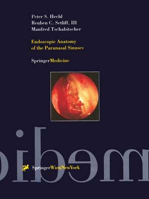
Endoscopic Anatomy of the Paranasal Sinuses
Autor Peter S. Hechl, Reuben C., III Setliff, Manfred Tschabitscheren Limba Engleză Paperback – 7 noi 2012
| Toate formatele și edițiile | Preț | Express |
|---|---|---|
| Paperback (1) | 1407.66 lei 6-8 săpt. | |
| SPRINGER VIENNA – 7 noi 2012 | 1407.66 lei 6-8 săpt. | |
| Hardback (1) | 1308.43 lei 38-44 zile | |
| SPRINGER VIENNA – 27 mai 1997 | 1308.43 lei 38-44 zile |
Preț: 1407.66 lei
Preț vechi: 1481.75 lei
-5% Nou
Puncte Express: 2111
Preț estimativ în valută:
269.35€ • 293.50$ • 226.97£
269.35€ • 293.50$ • 226.97£
Carte tipărită la comandă
Livrare economică 23 aprilie-07 mai
Preluare comenzi: 021 569.72.76
Specificații
ISBN-13: 9783709173459
ISBN-10: 3709173450
Pagini: 135
Ilustrații: XV, 135 p.
Dimensiuni: 210 x 280 x 20 mm
Greutate: 0.37 kg
Ediția:Softcover reprint of the original 1st ed. 1997
Editura: SPRINGER VIENNA
Colecția Springer
Locul publicării:Vienna, Austria
ISBN-10: 3709173450
Pagini: 135
Ilustrații: XV, 135 p.
Dimensiuni: 210 x 280 x 20 mm
Greutate: 0.37 kg
Ediția:Softcover reprint of the original 1st ed. 1997
Editura: SPRINGER VIENNA
Colecția Springer
Locul publicării:Vienna, Austria
V-ar putea interesa
-
 Bailey's Head and Neck Surgery - Otolaryngology ReviewClark A. Rosen MD, FACS-13%Preț: 533.12 lei610.19 lei
 -
 -5%Preț: 517.98 lei545.23 lei
 -
 Cochlear ImplantsS Waltzman-23%Preț: 535.37 lei695.29 lei
 -
 Paranasal Sinuses: Anatomy, Development & Biological FunctionRamzi Tamer Younis-14%Preț: 1594.98 lei1852.86 lei
 -
 Obstructive & Central Sleep ApneaJoan Mccormick-14%Preț: 757.12 lei881.33 lei
 -
 Pediatric ENTJohn M. Graham-5%Preț: 1132.87 lei1192.49 lei
 -
 Rhinology and Facial Plastic SurgeryFred J. Stucker-5%Preț: 1462.76 lei1539.74 lei
 -
 A Practical Guide to the Eustachian TubeJohn L. Dornhoffer-5%Preț: 516.04 lei543.21 lei
 -
 Vertigo and Balance Disorders in ChildrenKimitaka Kaga-5%Preț: 714.46 lei752.06 lei
 -
 Regenerative Medicine for the Inner EarJuichi Ito-11%Preț: 1033.70 lei1161.47 lei
Public țintă
ResearchCuprins
1. The nasal septum.- 2. The ethmoid bone and middle turbinate.- 3. The middle meatus.- 4. The uncinate process.- 5. The hiatus semilunaris and infundibulum.- 6. The ethmoidal bulla.- 7. The basal lamella.- 8. The maxillary sinus ostium, final common pathway, and exit of infundibulum.- 9. The sinus lateralis.- 10. The agger nasi cell.- 11. The Haller cell.- 12. The maxillary sinus.- 13. The posterior ethmoid.- 14. The superior turbinate.- 15. The sphenoid sinus.- 16. The skull base (ribbed vault).- 17. The frontal sinus.
Recenzii
"... an atlas of surgical endonasal anatomy comprising exhaustive illustrations of very high quality. The legends are clearly appended ... The colors and contrasts which are important aids in endoscopy are perfectly presented ... this atlas appears very useful for understanding the anatomy of the ethmoidal labyrinth. It will familiarise the surgeon’s eye with the fundamental anatomic landmarks in normal and pathologic conditions ...” Surgical and Radiologic Anatomy"... an important aid to the acquisition of endoscopic knowledge and ability ...” Australian Journal of Otolaryngology"... a leading anatomical reference book for planning of endoscopic procedures involving the paranasal sinuses and therefore this book should prove useful to ENT-surgeons as well as to neurosurgeons ...” Minimally Invasive Neurosurgery