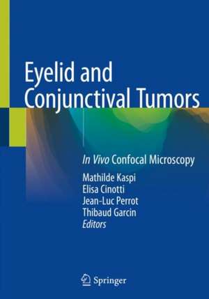
Eyelid and Conjunctival Tumors: In Vivo Confocal Microscopy
Editat de Mathilde Kaspi, Elisa Cinotti, Jean-Luc Perrot, Thibaud Garcinen Limba Engleză Paperback – 29 mar 2021
| Toate formatele și edițiile | Preț | Express |
|---|---|---|
| Paperback (1) | 734.04 lei 38-44 zile | |
| Springer International Publishing – 29 mar 2021 | 734.04 lei 38-44 zile | |
| Hardback (1) | 1023.52 lei 38-44 zile | |
| Springer International Publishing – 29 mar 2020 | 1023.52 lei 38-44 zile |
Preț: 734.04 lei
Preț vechi: 772.67 lei
-5% Nou
Puncte Express: 1101
Preț estimativ în valută:
140.48€ • 145.12$ • 116.91£
140.48€ • 145.12$ • 116.91£
Carte tipărită la comandă
Livrare economică 21-27 martie
Preluare comenzi: 021 569.72.76
Specificații
ISBN-13: 9783030366087
ISBN-10: 3030366081
Pagini: 134
Ilustrații: VIII, 134 p. 132 illus. in color.
Dimensiuni: 178 x 254 mm
Greutate: 0.32 kg
Ediția:1st ed. 2020
Editura: Springer International Publishing
Colecția Springer
Locul publicării:Cham, Switzerland
ISBN-10: 3030366081
Pagini: 134
Ilustrații: VIII, 134 p. 132 illus. in color.
Dimensiuni: 178 x 254 mm
Greutate: 0.32 kg
Ediția:1st ed. 2020
Editura: Springer International Publishing
Colecția Springer
Locul publicării:Cham, Switzerland
Cuprins
Preface.- I EXAMINATION OF THE OCULAR AND PERIOCULAR SURFACE.- 1 Clinical examination of the eyelid and conjunctiva.- 2 In vivo Reflectance Confocal Microscopy examination of eyelid and conjunctiva.- 3 Histopathological examination of the eyelid and conjunctiva.- II EYELID AND EYELID MARGIN.- 4 The normal eyelid.- 5 Benign epidermal lesions of the eyelid: Squamous papilloma.- 6 Benign epidermal lesions of the eyelid: Molluscum contagiosum.- 7 Benign epidermal lesions of the eyelid: Seborrheic keratosis.- 8 Benign epidermal lesions of the eyelid: Melanoacanthoma.- 9 Benign epidermal lesions of the eyelid: Epidermal cyst.- 10 Precancerous epidermal lesions of the eyelid: Actinic keratosis.- 11 Malignant epidermal tumors: Squamous cell carcinoma.- 12 Malignant epidermal tumors: Basal cell carcinoma.- 13 Benign lesions with basal melanocyte proliferation: Actinic lentigo.- 14 Benign melanocytic tumors: Junctional nevus.- 15 Benign melanocytic tumors: Subepithelial nevus.- 16 Benign melanocytic tumors: Compound nevus.- 17 Malignant melanocytic tumors : Melanoma.- 18 Benign Adnexal tumors of the eyelid: Trichoepitelioma.- 19 Benign Adnexal tumors of the eyelid: Pilomatricoma.- 20 Benign Adnexal tumors of the eyelid: Hidrocystoma.- 21 Malignant adnexal tumors of th eyelid: Sebaceous carcinoma.- 22 Vascular tumors of the eyelid: Infantile Hemangioma.- 23 Vascular tumors of the eyelid: Lobular capillary Hemangioma.- 24 Miscellaneous tumors of the eyelid: Nerve sheath tumors (Neurofibroma).- 25 Non lymphoid cutaneous infiltrates (Xanthelasma).- III CONJUNCTIVA.- 26 Normal conjunctiva.- 27 Benign conjunctival lesions: Pterygium.- 28 Benign conjunctival lesions: Primary acquired melanosis.- 29 Benign conjunctival lesions:: Nevus.- 30 Primary acquired melanosis: Epithelial cystic nevus.- 31 Malignant conjunctival tumors: Squamous cell carcinoma.- 32 Malignant conjunctival tumors: Melanoma.- 33 Malignant conjunctival tumors: B-cell Lymphoma.
Notă biografică
Mathilde Kaspi is Ophthalmologist (MD) at the Hospital of Montbrison and at University Hospital of Saint-Etienne. She is the principal author of this innovative work linking dermatology, ophthalmology, and pathology, focusing on the analysis of in vivo confocal microscopy (IVCM) images of various conjunctival and eye lid diseases, and in particular on their agreement with the histopathological diagnosis. As member of French society of Ophthalmologic Plastic Reconstructive Aesthetics, her work is focused on research, medical and surgical treatment of eye and eyelids pathologies and especially tumours.
Elisa Cinotti, PdD, Professor in Dermatology, graduated and did her Dermatology residency at the University of Genoa. From 2012 to 2016 she worked at the University Hospital of Saint-Etienne (France) and from 2016 she is working at the University Hospital of Siena (Italy) where she is conducting several researches on cutaneous and ocular noninvasive imaging, in particular on Reflectance Confocal Microscopy. She published over 150 peer-reviewed articles, most of them about dermatologic imaging.
Jean-Luc Perrot, PdD, Professor in Dermatology, graduated and did his Dermatology residency at the University of Saint-Etienne . From 1991 he has been working at the University Hospital of Saint-Etienne (France) where he is conducting several researches on cutaneous and ocular noninvasive imaging, in particular on Reflectance Confocal Microscopy. He has published over 200 peer-reviewed articles, most of them about dermatologic imaging. He is the president of the noninvasive cutaneous imaging group of the French Society of Dermatology.
Thibaud Garcin is Ophthalmologist (MD) at the University Hospital of Saint-Etienne and is currently fellow in vitreo-retinal surgery and anterior segment surgery. After MSc in Sciences and Health Engineering, he has supported his PhD (autumn 2019) about new device and new controls quality for corneal grafts in the laboratory ‘Biology engineering and imaging of Corneal Graft’. He is member of French and European societies for cornea and retina. His work is focused on medico-surgical treatment of the entire eye and research especially new techniques of imaging.
Textul de pe ultima copertă
This Atlas gives the complete expert opinion on the diagnostic features of eyelid and conjunctival tumors (benign and malignant): a state-of-the-art guide with numerous images, useful for both dermatologists and ophthalmologists. This invaluable resource, illustrating clinical, histological and re fectance confocal microscopy features, first addresses the normal conditions of the ocular surface, then reviews lesions due to epidermal, melanocytic and adnexal tumors. A final part is devoted to conjunctiva conditions, from normal to malignant conjunctival tumors. The high number of illustrations and their description of many ocular surface lesions with in vivo confocal microscopy make this atlas an essential guide for the practitioners of both specialities.
Caracteristici
Complete expert opinion on the diagnostic features of eyelid and conjunctival tumors Provides clinical, histological and reflectance confocal microscopy features State-of-the-art Atlas with a wealth of high quality illustrations
