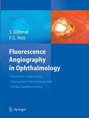
Fluorescence Angiography in Ophthalmology
Autor Stefan Dithmar, Frank G. Holzen Limba Engleză Paperback – 23 aug 2016
| Toate formatele și edițiile | Preț | Express |
|---|---|---|
| Paperback (1) | 859.02 lei 6-8 săpt. | |
| Springer Berlin, Heidelberg – 23 aug 2016 | 859.02 lei 6-8 săpt. | |
| Hardback (1) | 1185.87 lei 6-8 săpt. | |
| Springer Berlin, Heidelberg – 26 mar 2008 | 1185.87 lei 6-8 săpt. |
Preț: 859.02 lei
Preț vechi: 904.23 lei
-5% Nou
Puncte Express: 1289
Preț estimativ în valută:
164.37€ • 171.62$ • 136.04£
164.37€ • 171.62$ • 136.04£
Carte tipărită la comandă
Livrare economică 05-19 aprilie
Preluare comenzi: 021 569.72.76
Specificații
ISBN-13: 9783662517918
ISBN-10: 3662517914
Pagini: 224
Ilustrații: X, 224 p.
Dimensiuni: 210 x 279 mm
Greutate: 0.84 kg
Ediția:Softcover reprint of the original 1st ed. 2008
Editura: Springer Berlin, Heidelberg
Colecția Springer
Locul publicării:Berlin, Heidelberg, Germany
ISBN-10: 3662517914
Pagini: 224
Ilustrații: X, 224 p.
Dimensiuni: 210 x 279 mm
Greutate: 0.84 kg
Ediția:Softcover reprint of the original 1st ed. 2008
Editura: Springer Berlin, Heidelberg
Colecția Springer
Locul publicării:Berlin, Heidelberg, Germany
Cuprins
The physical and chemical fundamentals of fluorescence angiography.- The Technical Fundamentals of Fluorescence Angiography.- Normal Fluorescence Angiography and General Pathological Fluorescence Phenomena.- Fundus Autofluorescence.- Macular Disorders.- Retinal Vascular Disease.- Inflammatory Retinal/Choroidal Disease.- Diseases of the Optic Nerve Head.- Intraocular Tumors.
Recenzii
From the reviews:“This excellent new angiography atlas by two German ophthalmologists has been translated into English and I suspect will quickly find its way into the libraries of UK ophthalmology departments. … The book reads very well indeed. … The layout works very well, and I have found it a very helpful book to refer to when looking at angiograms and autofluorescence images from my own patients. … the absolute key to a successful atlas in clinical ophthalmology is image quality.” (James Cameron, Eye News, February/March, 2010)
Textul de pe ultima copertă
The technology of angiographic systems has been improved tremendously just within the past few years. This has allowed greatly increased levels of image resolution for both fluorescein and indocyanine green angiography.
This new atlas by Dithmar and Holz covers the basic principles of the new methods for fluorescein- and indocyanine green-angiography, as well as the high resolution imaging of fundus autofluorescence.
The angiographic signs of retinal and choroidal diseases are illustrated with images taken from a series of clinically relevant case examples that specifically illustrate the advantages of higher image resolution for the study of common retinochoroidal disorders. In so doing, this atlas offers an all-encompassing survey of the many angiographic signs in these disorders and their differential diagnoses. Clinicians in all subspecialties of ophthalmology can profit from a better understanding of these pathophysiological phenomena.
