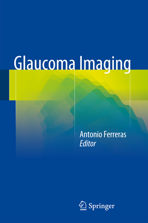
Glaucoma Imaging
Editat de Antonio Ferrerasen Limba Engleză Hardback – 30 oct 2015
| Toate formatele și edițiile | Preț | Express |
|---|---|---|
| Paperback (1) | 1033.37 lei 6-8 săpt. | |
| Springer International Publishing – 23 aug 2016 | 1033.37 lei 6-8 săpt. | |
| Hardback (1) | 1172.33 lei 3-5 săpt. | |
| Springer International Publishing – 30 oct 2015 | 1172.33 lei 3-5 săpt. |
Preț: 1172.33 lei
Preț vechi: 1234.02 lei
-5% Nou
Puncte Express: 1758
Preț estimativ în valută:
224.40€ • 243.83$ • 188.61£
224.40€ • 243.83$ • 188.61£
Carte disponibilă
Livrare economică 31 martie-14 aprilie
Preluare comenzi: 021 569.72.76
Specificații
ISBN-13: 9783319189581
ISBN-10: 3319189581
Pagini: 250
Ilustrații: VI, 328 p.
Dimensiuni: 155 x 235 x 22 mm
Greutate: 0.79 kg
Ediția:1st ed. 2016
Editura: Springer International Publishing
Colecția Springer
Locul publicării:Cham, Switzerland
ISBN-10: 3319189581
Pagini: 250
Ilustrații: VI, 328 p.
Dimensiuni: 155 x 235 x 22 mm
Greutate: 0.79 kg
Ediția:1st ed. 2016
Editura: Springer International Publishing
Colecția Springer
Locul publicării:Cham, Switzerland
Public țintă
Professional/practitionerCuprins
Standard Automated Perimetry.- Gonioscopy.- Measurement of Intraocular Pressure.- Clinical Applications of Ultrasound Biomicroscopy in Glaucoma.- Retrobulbar Ocular Blood Flow Evaluation in Open Angle Glaucoma.- Optic Nerve Head Assessment and Retinal Nerve Fiber Layer Evaluation.- Confocal Scanning Laser Ophthalmoscopy (HRT).- Detection of Glaucoma Using Scanning Laser Polarimetry.- Optical Coherence Tomography (OCT).- Measuring Hemoglobin Levels in the Optic Nerve Head for Glaucoma Management.- Structure & Function Relationship in Glaucoma.- Assessment of Structural Glaucoma Progression.
Recenzii
“The purpose is to provide an update on the use ofimaging technologies for glaucoma. This is a good resource forophthalmologists, ophthalmology residents, optometrists, and glaucomaresearchers. … This is a valuable resource for clinicians and researchers tohelp improve the quality of care for glaucoma patients.” (Diana V. Do, Doody’sBook Reviews, February, 2016)
Notă biografică
Antonio Ferreras, MD, PhD, is a Consultant Ophthalmologist who works at the Miguel Servet University Hospital, Zaragoza, Spain and the Aragon Health Sciences Institute, University of Zaragoza. He has been an Associate Professor at the University of Zaragoza since 2007. Until 2010 he worked as a glaucoma and cataract surgeon, and he is currently a vitreoretinal and cataract surgeon. Dr. Ferreras has been involved in numerous clinical trials and his research has included the diagnosis of glaucoma by means of high-definition optical coherence tomography and scanning laser polarimetry with enhanced corneal compensation. He is the recipient of an International Ophthalmologist Education Award (2009) and an Achievement Award (2013) from the American Academy of Ophthalmology. Dr. Ferreras is a member of the editorial board of Case Reports in Ophthalmological Medicine and a member of the International Journal of Ophthalmology Expert Board. He has been guest editor for a Journal of Ophthalmology special edition on “Different aspects in different glaucomas” (2010-11) and for the BioMed Research International special issue on “Advances in imaging technologies for evaluating the retina and the optic disc” (2014). He is the author of more than 80 peer-reviewed publications.
Textul de pe ultima copertă
This atlas offers a truly comprehensive update on the use of imaging technologies for the diagnosis and follow-up of glaucoma. In addition to standard automated perimetry, gonioscopy, and fundus photography, other advanced high-resolution methods for imaging the eye in glaucoma are explained in detail, including ultrasound biomicroscopy, confocal scanning laser ophthalmoscopy, scanning laser polarimetry, and spectral domain optical coherence tomography. The role of the various tests and the keys to optimizing their use in clinical practice are detailed with the aid of high-quality figures in order to enable the reader to achieve the best possible performance when applying these tools.
The risk of developing visual disability and blindness as a consequence of glaucoma varies widely among affected individuals. Personalized testing strategies and tailored therapeutic interventions are required to effectively reduce visual impairment due to glaucoma. Glaucoma Imagingwill assist residents, researchers, and clinicians in improving their ability to understand and integrate the information obtained using traditional techniques with the reports provided by computer-assisted image instruments.
The risk of developing visual disability and blindness as a consequence of glaucoma varies widely among affected individuals. Personalized testing strategies and tailored therapeutic interventions are required to effectively reduce visual impairment due to glaucoma. Glaucoma Imagingwill assist residents, researchers, and clinicians in improving their ability to understand and integrate the information obtained using traditional techniques with the reports provided by computer-assisted image instruments.
Caracteristici
Offers comprehensive guidance on use of imaging technologies for diagnosis and follow-up Explains the roles of both traditional and advanced imaging techniques Identifies the keys to optimizing use of the different methods
