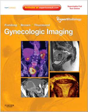
Gynecologic Imaging: Expert Radiology Series (Expert Consult Premium Edition - Enhanced Online Features and Print): Expert Radiology
Autor Julia R. Fielding, Douglas L. Brown, Amy S. Thurmonden Limba Engleză Hardback – 13 mai 2011
Preț: 1206.42 lei
Preț vechi: 1558.68 lei
-23% Nou
Puncte Express: 1810
Preț estimativ în valută:
230.84€ • 241.67$ • 191.01£
230.84€ • 241.67$ • 191.01£
Carte disponibilă
Livrare economică 21 martie-02 aprilie
Preluare comenzi: 021 569.72.76
Specificații
ISBN-13: 9781437715750
ISBN-10: 1437715753
Pagini: 688
Ilustrații: Approx. 1200 illustrations (245 in full color)
Dimensiuni: 222 x 281 x 35 mm
Greutate: 0 kg
Ediția:New.
Editura: Elsevier
Seria Expert Radiology
ISBN-10: 1437715753
Pagini: 688
Ilustrații: Approx. 1200 illustrations (245 in full color)
Dimensiuni: 222 x 281 x 35 mm
Greutate: 0 kg
Ediția:New.
Editura: Elsevier
Seria Expert Radiology
Cuprins
1.
The
Normal
Pelvis
on
Ultrasound
imaging
and
Anatomical
Correlations
2. PITFALLS IN GYNECOLOGIC ULTRASOUND
3. CT - Normal anatomy, Imaging techniques and pitfalls
4. CT - Dose reduction Techniques in MDCT Body Imaging
5. Common protocols
6. New techniques, diffusion
7. Hysterosalpingography: Techniques, Normal Anatomy, and Pitfalls
8. Approach to Pelvic Pain and the Role of Imaging
9. Endometriosis
10. Acute Pelvic Pain: An Overview
11. Chronic Pelvic Pain
12. Pelvic Pain: Lower Urinary Tract - Urethral Diverticulum, Cysts, Varix
13. Benign Endometrial Causes of Abnormal Bleeding
14. Adenomyosis
15. Uterine Leiomyomas
16. Overview
17. Tubal abnormalities
18. Mullerian uterine anomalies
19. The Ovary and Polycystic Ovary Syndrome
20. THE IMAGING OF CONTRACEPTION
21. Ultrasound of the Normal and Failed First Trimester Pregnancy
22. Ectopic Pregnancy
23. Retained products of conception
24. Gestational Trophoblastic Neoplasia
25. Postpartum-retained placenta, post C-section, etc.
26. Vaginal fistulas
27. Pelvic prolapsed
28. Approach to Imaging the adnexal mass
29. Benign ovarian masses
30. Malignant ovarian masses
31. Use of PET imaging in gynecologic cancers
32. Uterine cancers
33. Cervical cancer
34. Ovarian cancer/fallopian tube cancer
35. Carcinoma of the Vagina and Vulva
36. Gynecologic imaging of the pediatric patient
37. Drainage and Biopsy Procedures
38. Focused Ultrasound Ablation of Uterine Leiomyomas
39. UAE
40. Fallopian tube catheterization
41. Ultrasound Guided Treatment of ectopic pregnancy
