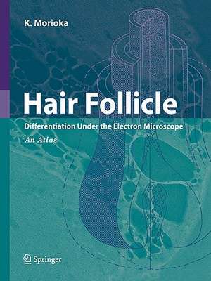
Hair Follicle: Differentiation under the Electron Microscope - An Atlas
Autor K. Moriokaen Limba Engleză Paperback – 20 oct 2010
| Toate formatele și edițiile | Preț | Express |
|---|---|---|
| Paperback (1) | 1293.41 lei 38-44 zile | |
| Springer – 20 oct 2010 | 1293.41 lei 38-44 zile | |
| Hardback (1) | 1316.76 lei 38-44 zile | |
| Springer – 13 dec 2004 | 1316.76 lei 38-44 zile |
Preț: 1293.41 lei
Preț vechi: 1361.48 lei
-5% Nou
Puncte Express: 1940
Preț estimativ în valută:
247.57€ • 269.01$ • 208.10£
247.57€ • 269.01$ • 208.10£
Carte tipărită la comandă
Livrare economică 17-23 aprilie
Preluare comenzi: 021 569.72.76
Specificații
ISBN-13: 9784431998051
ISBN-10: 4431998055
Pagini: 164
Ilustrații: XIII, 152 p. 89 illus., 6 illus. in color.
Dimensiuni: 210 x 277 x 9 mm
Greutate: 0.39 kg
Ediția:Softcover reprint of hardcover 1st ed. 2005
Editura: Springer
Colecția Springer
Locul publicării:Tokyo, Japan
ISBN-10: 4431998055
Pagini: 164
Ilustrații: XIII, 152 p. 89 illus., 6 illus. in color.
Dimensiuni: 210 x 277 x 9 mm
Greutate: 0.39 kg
Ediția:Softcover reprint of hardcover 1st ed. 2005
Editura: Springer
Colecția Springer
Locul publicării:Tokyo, Japan
Public țintă
ResearchDescriere
Each and every hair is much more than just the visible shaft—there are also associated complex sheath structures of epidermal and dermal origin. In the hair follicle, cells undergo a variety of differentiation processes, mostly depending on their layers and positions therein, and electron microscopy reveals a very complex architecture. The structure of a particular layer, such as Henle’s layer of the inner root sheath, is not uniform. Rather, cells drastically change during the course of differentiation. By simply comparing electron micrographs of cells of a layer at different degrees of differentiation, one can hardly recognize them as belonging to the same layer. As readers will see, this book contains many superb electron mic- graphs, from low-magni?cation panoramic views for orientation to hi- power views showing ultrastructural detail. Captions and schematic drawings are also very helpful in “reading” electron micrographs and - derstanding the structural detail. In this way, Dr. Morioka has succeeded in dissecting the complex hair follicle at the ultrastructural level.
Cuprins
Medulla.- Hair Cortex and Hair Cuticle.- Inner Root Sheath.- Outer Root Sheath and Companion Layer.- Hair Bulb and Papilla.
Caracteristici
Provides a quick understanding of the structure and development of hair follicle with a lot of clear electron microscope photographs
