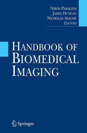
Handbook of Biomedical Imaging: Methodologies and Clinical Research
Editat de Nikos Paragios, James Duncan, Nicholas Ayacheen Limba Engleză Hardback – 25 mar 2015
The dominant state-of-the-art methodologies for content extraction and interpretation of medical images include fuzzy methods, level set methods, kernel methods, and geometric deformable models. The models and techniques discussed are used in the diagnosis, planning, control and follow-up of medical procedures. Throughout the book, challenges and limitations are explored along with new research directions.
Handbook of Biomedical Imaging offers a unique guide to the entire chain of biomedical imaging, explaining how image formation is done and how the most appropriate techniques are used to address demands and diagnoses. Radiologists, research scientists, senior undergraduate and graduate students in health sciences and engineering, and university professors will find this to be an exceptional reference.
| Toate formatele și edițiile | Preț | Express |
|---|---|---|
| Paperback (1) | 1178.75 lei 38-44 zile | |
| Springer Us – 5 oct 2016 | 1178.75 lei 38-44 zile | |
| Hardback (1) | 1289.09 lei 6-8 săpt. | |
| Springer Us – 25 mar 2015 | 1289.09 lei 6-8 săpt. |
Preț: 1289.09 lei
Preț vechi: 1611.36 lei
-20% Nou
Puncte Express: 1934
Preț estimativ în valută:
246.66€ • 257.56$ • 203.69£
246.66€ • 257.56$ • 203.69£
Carte tipărită la comandă
Livrare economică 15-29 aprilie
Preluare comenzi: 021 569.72.76
Specificații
ISBN-13: 9780387097480
ISBN-10: 0387097481
Pagini: 511
Ilustrații: VII, 511 p. 154 illus.
Dimensiuni: 155 x 235 x 29 mm
Greutate: 0.9 kg
Ediția:2015
Editura: Springer Us
Colecția Springer
Locul publicării:New York, NY, United States
ISBN-10: 0387097481
Pagini: 511
Ilustrații: VII, 511 p. 154 illus.
Dimensiuni: 155 x 235 x 29 mm
Greutate: 0.9 kg
Ediția:2015
Editura: Springer Us
Colecția Springer
Locul publicării:New York, NY, United States
Public țintă
ResearchCuprins
Object Segmentation and Markov Random Fields.- Fuzzy methods in medical imaging.- Curve Propagation, Level Set Methods and Grouping.- Kernel Methods in Medical Imaging.- Geometric Deformable Models: Overview and Recent Developments.- Active Shape and Appearance Models.- Statistical Atlases.- Statistical Computing on Non-Linear Spaces for Computational Anatomy.- Building Patient-Specific Physical and Physiological Computational Models from Medical Images.- Constructing a Patient-Specific Model Heart from CT Data.- Image-based haemodynamics simulation in intracranial aneursyms.- Atlas-based Segmentation.- Integration of Topological Constraints in Medical Image Segmentation.- Monte Carlo Sampling for the Segmentation of Tubular Structures.- Non-rigid registration using free-form deformations.- Image registration using mutual information.- Physical Model Based Recovery of Displacement and Deformations from 3D medical images.- Cardiovascular Informatics.- Rheumatoid Arthritis Quantifiction using Appearance Models.- Medical Image Processing for Analysis of Colon Motility.- Segmentation of Diseased Livers: A 3D Refinement Approach.- Intra and inter subject analyses of brain functional Magnetic Resonance Images (fMRI).- Diffusion Tensor Estimation, Regularization and Classification.- From Local Q-Ball Estimation to Fibre Crossing Tractography.- Segmentation of clustered cells in microscopy images by geometric PDEs and level sets.- Atlas-based whole-body registration in mice.
Notă biografică
Nikos Paragios (D.Sc. (2005), PhD (2000), M.Sc. (1996), B.Sc. (1994)) is professor of Applied Mathematics, director of the Center for Visual Computing of Ecole Centrale de Paris, France and scientific leader of GALEN project-team of Ecole Centrale de Paris/Inria Saclay, Ile-de-France. Professor Paragios is an IEEE Fellow (2012), has co-edited four books, published more than two hundred fifty papers in the most prestigious journals and conferences of medical imaging and computer vision, and holds twenty one international patents (http://cvn.ecp.fr/personnel/nikos/). Professor Paragios is the Editor in Chief of the Computer Vision and Image Understanding Journal (CVIU), member of the editorial board of the the Medical Image Analysis Journal(MedIA) and the SIAM Journal in Imaging Sciences (SIIMS) , while has served as an associate/area editor/member of the editorial board for the IEEE Transactions on Pattern Analysis and Machine Intelligence (PAMI), the International Journal of Computer Vision (IJCV), the Journal of Mathematical Imaging and Vision (JMIV), the Imaging and Vision Computing Journal (IVC), the Machine Vision and Applications (MVA) Journal. Professor Paragios serves regularly at the conference boards of the most prestigious events of his field (ICCV, CVPR, ECCV, MICCAI) while being member of the scientific council of SAFRAN conglomerate.
Nicholas Ayache is a Research Director at INRIA, France, and the scientific leader of the ASCLEPIOS project-team in Sophia Antipolis. His current research interests include the analysis and simulation of biomedical images with advanced geometrical, statistical,biophysical and functional models, and the application of these tools to medicine to improve the diagnosis and therapy of diseases. His publications, which have received over 25,000 citations (according to scholar), may be found at: http://www-sop.inria.fr/members/Nicholas.Ayache. Dr. Ayache received in 2008 the "Microsoft Award for Science in Europe"awarded jointly by the Royal Society and the French Academy of Sciences. In 2013 he received in Nagoya (Japan) the 2013 Miccai Enduring Impact Award. For the academic year 2013-2014, Dr. Ayache was elected a professor at the Collège de France, on the Chair Informatics and Digital Sciences. Dr. Ayache has been a scientific consultant for several companies and international research institutes, and has been a co-founder of several start-up companies in image processing, computer vision, and biomedical imaging. Dr. Ayache has been a founding Editor of the Medical Image Analysis Journal (Elsevier Science), an associate Editor of IEEE Trans. on Medical Imaging, and the general Chair of the MICCAI 2012 conference in Nice.
James S. Duncan is the Ebenezer K. Hunt Professor of Biomedical Engineering and a Professor of Diagnostic Radiology and Electrical Engineering at Yale University. Professor Duncan received his B.S.E.E. with honors from Lafayette College (1973), his M.S. (1975) from UCLA and Ph.D. (1982) in Electrical Engineering from the University of Southern California. Professor Duncan has been at Yale University since 1983, and the Ebenezer K. Hunt Professor of Biomedical Engineering at Yale University since 2007. He has served as the Acting Chair and is currently Director of Undergraduate Studies for Biomedical Engineering. His research efforts have been in the areas of computer vision, image processing, and medical imaging, with an emphasis on biomedical image analysis. These efforts have included the segmentation of deformable structure from 3D image data, the tracking of non-rigid motion/deformation from spatiotemporal images, and the development of strategies for image-guided intervention/surgery. He has published over 220 peer-reviewed articles in these areas and has been the principal investigator on a number of peer-reviewed grants from both the National Institutes of Health and the National ScienceFoundation over the past 28 years. Professor Duncan is a Fellow of the Institute of Electrical and Electronic Engineers (IEEE), of the American Institute for Medical and Biological Engineering (AIMBE) and of the Medical Image Computing and Computer Assisted Intervention (MICCAI) Society. He currently serves as co-Editor-in-Chief of Medical Image Analysis and as an Associate Editor of IEEE Transactions on Medical Imaging. In 2012, he was elected to the Council of Distinguished Investigators, Academy of Radiology Research. In 2014, he was elected to the Connecticut Academy of Science and Engineering.
Textul de pe ultima copertă
Biomedical image analysis has become a major aspect of engineering sciences, and radiology in particular has become a dominant player in the field. Recent developments have made it possible to use biomedical imaging to view the human body from an anatomical or physiological perspective in a non-invasive fashion. Computer-aided diagnosis consists of developing algorithms and intelligent software components that can automatically process images and spot potential irregularities in the health chain.
This book explains the process of computer assisted biomedical image analysis diagnosis through mathematical modeling and inference of image-based bio-markers. It covers five crucial thematic areas: methodologies, statistical and physiological models, biomedical perception, clinical biomarkers, and emerging modalities and domains.
The dominant state-of-the-art methodologies for content extraction and interpretation of medical images include fuzzy methods, level set methods, kernel methods, and geometric deformable models. The models and techniques discussed are used in the diagnosis, planning, control and follow-up of medical procedures. Throughout the book, challenges and limitations are explored along with new research directions.
This complete volume is an exceptional tool for radiologists, research scientists, senior undergraduate and graduate students in health sciences and engineering, and university professors. This book offers a unique guide to the entire chain of biomedical imaging, explaining how image formation is done, and how the most appropriate algorithms are used to address demands and diagnoses.
Caracteristici
Authors are leaders in the field Current and thorough review of biomedical image analysis Application-oriented Authors from both Computer Science and Radiology communities


