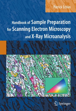
Handbook of Sample Preparation for Scanning Electron Microscopy and X-Ray Microanalysis
Autor Patrick Echlinen Limba Engleză Hardback – 19 mar 2009
| Toate formatele și edițiile | Preț | Express |
|---|---|---|
| Paperback (1) | 744.86 lei 38-44 zile | |
| Springer Us – 23 noi 2010 | 744.86 lei 38-44 zile | |
| Hardback (1) | 901.71 lei 6-8 săpt. | |
| Springer Us – 19 mar 2009 | 901.71 lei 6-8 săpt. |
Preț: 901.71 lei
Preț vechi: 1099.64 lei
-18% Nou
Puncte Express: 1353
Preț estimativ în valută:
172.54€ • 180.63$ • 142.77£
172.54€ • 180.63$ • 142.77£
Carte tipărită la comandă
Livrare economică 05-19 aprilie
Preluare comenzi: 021 569.72.76
Specificații
ISBN-13: 9780387857305
ISBN-10: 0387857303
Pagini: 330
Ilustrații: XII, 332 p. 159 illus. in color.
Dimensiuni: 178 x 254 x 23 mm
Greutate: 0.91 kg
Ediția:2009
Editura: Springer Us
Colecția Springer
Locul publicării:New York, NY, United States
ISBN-10: 0387857303
Pagini: 330
Ilustrații: XII, 332 p. 159 illus. in color.
Dimensiuni: 178 x 254 x 23 mm
Greutate: 0.91 kg
Ediția:2009
Editura: Springer Us
Colecția Springer
Locul publicării:New York, NY, United States
Public țintă
ResearchDescriere
Scanning electr on microscopy (SEM) and x-ray microanalysis can produce magnified images and in situ chemical information from virtually any type of specimen. The two instruments generally operate in a high vacuum and a very dry environment in order to produce the high energy beam of electrons needed for imaging and analysis. With a few notable exceptions, most specimens destined for study in the SEM are poor conductors and composed of beam sensitive light elements containing variable amounts of water. In the SEM, the imaging system depends on the specimen being sufficiently electrically conductive to ensure that the bulk of the incoming electrons go to ground. The formation of the image depends on collecting the different signals that are scattered as a consequence of the high energy beam interacting with the sample. Backscattered electrons and secondary electrons are generated within the primary beam-sample interactive volume and are the two principal signals used to form images. The backscattered electron coefficient ( ? ) increases with increasing atomic number of the specimen, whereas the secondary electron coefficient ( ? ) is relatively insensitive to atomic number. This fundamental diff- ence in the two signals can have an important effect on the way samples may need to be prepared. The analytical system depends on collecting the x-ray photons that are generated within the sample as a consequence of interaction with the same high energy beam of primary electrons used to produce images.
Cuprins
Sample Collection and Selection.- Sample Preparation Tools.- Sample Support.- Sample Embedding ?and Mounting.- Sample Exposure.- Sample Dehydration.- Sample Stabilization for Imaging in the SEM.- Sample Stabilization to Preserve Chemical Identity.- Sample Cleaning.- Sample Surface Charge Elimination.- Sample Artifacts and Damage.- Additional Sources of Information.
Recenzii
This handbook should find its way to the reference bookshelf of all imaging laboratories. It should also become required reading for anyone being trained for SEM work, or anyone who might need to have their samples examined by using such techniques. In that way, it will be less likely that deficient results will be published and that the full potential of the SEM be realized.-- Iolo ap Gwynn, Microscopy and Microanalysis (2010)
Notă biografică
Patrick Echlin was a lecturer in the Department of Plant Sciences and Director of the Multi-Imaging Centre, School of Biological Science, University of Cambridge until he retired in 1999. He has taught for more than thirty years at the Lehigh University Microscopy School and is the author and co-auther of eight books on scanning electron microscopy and x-ray microanalysis. He is an Honorary Fellow of the Royal Microscopical Society and received the Distinguished Scientist Award in Biological Sciences from the Microscope Society of America in 2001.
Textul de pe ultima copertă
This Handbook is a complete guide to preparing a wide variety of specimens for the scanning electron microscope and x-ray microanalyzers. Specimens range from inorganic, organic, biological, and geological samples to materials such as metals, polymers, and semiconductors which can exist as solids, liquids and gases. While the Handbook complements the best-selling textbook, Scanning Electron Microscopy and X-Ray Microanalysis, Third Edition, by Goldstein, et al., it is entirely self-contained and describes what is needed up to the point the sample is put into the instrument. Photomicrographs of each specimen complement the many sample preparation "recipes." Additional chapters describe the general features of specimen preparation in relation to the different needs of scanning electron microscopes and x-ray microanalyzers, and an appendix covers chemicals and equipment applicable to any of the recipes. This practical Handbook is an essential reference for anyone who uses these instruments. It assumes only an elementary knowledge of preparation techniques but also serves as an authoritative guide for more experienced microscopists.
Caracteristici
Identifies problems that all specimens present in examining their structure and analysis in the SEM
Describes a series of protocols to ensure that a specimen is properly prepared once the particular problems are identified
Guides the reader through a general approach to the problem before a particular procedure is applied to the requirements of a given sample
Designed both as an introduction for the novice and as a guide for the practicing scanning microscopist
