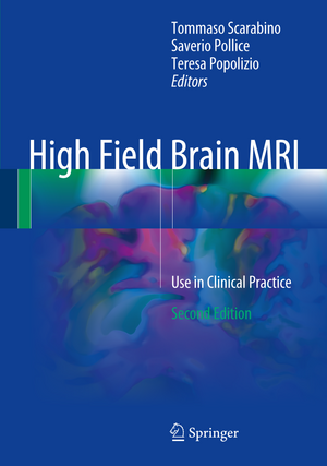
High Field Brain MRI: Use in Clinical Practice
Editat de Tommaso Scarabino, Saverio Pollice, Teresa Popolizioen Limba Engleză Hardback – 7 mar 2017
Preț: 867.20 lei
Preț vechi: 912.84 lei
-5% Nou
Puncte Express: 1301
Preț estimativ în valută:
165.96€ • 172.62$ • 137.01£
165.96€ • 172.62$ • 137.01£
Carte disponibilă
Livrare economică 25 martie-08 aprilie
Preluare comenzi: 021 569.72.76
Specificații
ISBN-13: 9783319441733
ISBN-10: 3319441736
Pagini: 391
Ilustrații: IX, 391 p. 175 illus., 82 illus. in color.
Dimensiuni: 178 x 254 x 29 mm
Greutate: 1.05 kg
Ediția:2nd ed. 2017
Editura: Springer International Publishing
Colecția Springer
Locul publicării:Cham, Switzerland
ISBN-10: 3319441736
Pagini: 391
Ilustrații: IX, 391 p. 175 illus., 82 illus. in color.
Dimensiuni: 178 x 254 x 29 mm
Greutate: 1.05 kg
Ediția:2nd ed. 2017
Editura: Springer International Publishing
Colecția Springer
Locul publicării:Cham, Switzerland
Cuprins
Techniques and Semeiotics.- High-Field MRI and Safety: I. Installation.- High-Field MRI and Safety: II. Utilization.- 3.0 T MRI Diagnostic Features: Comparison with Lower Magnetic Fields.- Standard 3.0 T MR Imaging.- 3.0 T MR Angiography.- 3.0 T MR Spectroscopy.- 3.0 T Diffusion Studies.- Nerve Pathways with MR Tractography.- 3.0 T Perfusion Studies.- High-Field Strength Functional MRI.- Recent Developments and Prospects in High-Field MR.- 3.0 T Brain MRI: A Pictorial Overview of the Most Interesting Sequences.- Applications.- High-Field Neuroimaging in Traumatic Brain Injury.- 3.0 T Imaging of Ischaemic Stroke.- High-Field Strength MRI (3.0 T or More) in White Matter Diseases.- High-Field Neuroimaging in Parkinson’s Disease.- High-Field 3 T Imaging of Alzheimer Disease.- 3.0 T Imaging of Brain Tumours.- Use of fMRI Activation Paradigms: A Presurgical Tool for Mapping Brain Function.
Notă biografică
Tommaso Scarabino, MD, currently works at the Radiology/Neuroradiology Department of Andria Hospital, Andria, Italy. Dr. Scarabino has edited 18 previous books and is the author of 80 scientific papers in Italian and international journals as well as 120 book chapters. He has been the scientific coordinator for more than 20 important meetings in Italy and is a regular invited speaker at courses and meetings.
Saverio Pollice, MD, currently works at the Radiology/Neuroradiology Department of Andria Hospital, Andria, Italy. He is the editor of one previous book and co-editor of a further six. He has also published scientific papers in Italian and international journals. Dr. Pollice has been an invited speaker at 20 courses and meetings.
Teresa Popolizio, MD, currently works at the Radiology/Neuroradiology Department of San Giovanni Rotondo Hospital in Italy. To date she has authored 70 scientific papers in Italian and international journals, as wellas 20 book chapters. She has also co-edited one previous book. Dr. Popolizio has been scientific coordinator for six important meetings in Italy and has been an invited speaker at numerous courses and meetings.
Textul de pe ultima copertă
This richly illustrated book summarizes the state of the art in brain MRI with the 3-Tesla high-field scanner. The aim is to clarify the numerous advantages of using a 3-T high magnetic field MR scanner, especially in terms of sensitivity and specificity. The first section describes techniques for standard MR examination of the brain. Full descriptions are then provided of advanced protocols such as MR angiography, diffusion-weighted imaging (DWI), perfusion-weighted imaging (PWI), MR spectroscopy, MR tractography, and functional imaging. Differences in diagnostic features when performing these examinations at 3 T and at 1.5 T are highlighted. In addition, safety issues relating to the installation and use of a high-field scanner are discussed. The second section then describes and illustrates in detail each of the main clinical applications of 3-T MRI in the human brain: trauma, stroke, white matter disease, Parkinson’s disease, Alzheimer’s disease, brain tumors, inflammatory disease, psychiatric disorders, etc. A final chapter is dedicated to the evaluation of 7-T MRI within both research and potential clinical settings. The book will be a valuable tool for general radiologists, neuroradiologists, trainees, and technicians.
Caracteristici
Offers an easy-to-use reference for radiologists and neuroradiologists working with high field strength MR scanners Describes the various MR techniques in detail Documents imaging findings in each of the main clinical applications of 3-T MRI Discusses safety issues relating to the installation and use of high-field magnets
