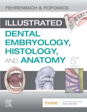
Illustrated Dental Embryology, Histology, and Anatomy
Autor Margaret J. Fehrenbach, Tracy Popowicsen Limba Engleză Paperback – 5 feb 2020
Preț: 466.65 lei
Preț vechi: 612.90 lei
-24% Nou
89.29€ • 93.23$ • 73.90£
Carte disponibilă
Livrare economică 08-22 martie
Livrare express 01-07 martie pentru 170.35 lei
Specificații
ISBN-10: 0323611079
Pagini: 352
Ilustrații: Approx. 685 illustrations (625 in full color)
Dimensiuni: 216 x 276 x 17 mm
Greutate: 0.95 kg
Ediția:5
Editura: Elsevier
Cuprins
UNIT I: OROFACIAL STRUCTURES 1. Face and Neck Regions 2. Oral Cavity and Pharynx
UNIT II: DENTAL EMBRYOLOGY 3. Prenatal Development 4. Face and Neck Development 5. Orofacial Development 6. Tooth Development and Eruption
UNIT III: DENTAL HISTOLOGY 7. Cells 8. Basic Tissue 9. Oral Mucosa 10. Gingival and Dentogingival Junctional Tissues 11. Head and Neck Structures 12. Enamel 13. Dentin and Pulp 14. Periodontium: Cementum, Alveolar Bone, Periodontal Ligament
UNIT IV: DENTAL ANATOMY 15. Overview of Dentitions 16. Permanent Anterior Teeth 17. Permanent Posterior Teeth 18. Primary Dentition 19. Temporomandibular Joint 20. Occlusion
Bibliography Glossary Appendix A: Anatomical Position Appendix B: Units of Measure Appendix C: Tooth Measurements Appendix D: Tooth Development Index
Descriere
Get a clear picture of oral biology and the formation and study of dental structures. Illustrated Dental Embryology, Histology, & Anatomy, 5th Edition is the ideal introduction to one of the most foundational areas in the dental professions - understanding the development, cellular makeup, and physical anatomy of the head and neck regions. Written in a clear, reader-friendly style, this text makes it easy for you to understand both basic science and clinical applications - putting the content into the context of everyday dental practice. New for the fifth edition is evidence-based research on the dental placode, nerve core region, bleeding difficulties, silver diamine fluoride, and primary dentition occlusion. Plus, high-quality color renderings and clinical histographs and photomicrographs throughout the book, truly brings the material to life.
