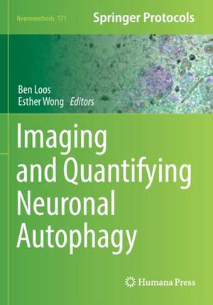
Imaging and Quantifying Neuronal Autophagy: Neuromethods, cartea 171
Editat de Ben Loos, Esther Wongen Limba Engleză Paperback – 21 aug 2022
Cutting-edge and practical, Imaging and Quantifying Neuronal is a valuable resource that provides insights into the power of microscopy tools, live-cell imaging, and photoactivation and correlative techniques.
| Toate formatele și edițiile | Preț | Express |
|---|---|---|
| Paperback (1) | 722.43 lei 6-8 săpt. | |
| Springer Us – 21 aug 2022 | 722.43 lei 6-8 săpt. | |
| Hardback (1) | 1115.46 lei 6-8 săpt. | |
| Springer Us – 20 aug 2021 | 1115.46 lei 6-8 săpt. |
Din seria Neuromethods
- 5%
 Preț: 347.57 lei
 - 15%
 Preț: 659.53 lei
 - 15%
 Preț: 665.08 lei
 - 18%
 Preț: 986.63 lei
 - 24%
 Preț: 852.89 lei
 - 18%
 Preț: 953.03 lei
 - 18%
 Preț: 955.25 lei
 - 20%
 Preț: 1129.36 lei
 - 20%
 Preț: 1252.04 lei
 - 18%
 Preț: 1291.45 lei
 - 15%
 Preț: 652.31 lei
 - 18%
 Preț: 955.70 lei
 - 23%
 Preț: 705.39 lei
 - 18%
 Preț: 973.38 lei
 - 18%
 Preț: 964.86 lei
 - 18%
 Preț: 968.03 lei
 - 15%
 Preț: 662.95 lei
 - 15%
 Preț: 646.43 lei
 - 15%
 Preț: 649.71 lei
 -
 Preț: 395.29 lei
 - 19%
 Preț: 580.67 lei
 - 19%
 Preț: 584.12 lei
 - 19%
 Preț: 566.41 lei
 - 15%
 Preț: 652.17 lei
 - 15%
 Preț: 655.13 lei
 - 18%
 Preț: 959.36 lei
 - 15%
 Preț: 652.49 lei
 - 15%
 Preț: 649.54 lei
 - 15%
 Preț: 649.87 lei
 - 15%
 Preț: 650.19 lei
 - 15%
 Preț: 648.42 lei
 - 18%
 Preț: 1039.22 lei
 - 18%
 Preț: 963.15 lei
Preț: 722.43 lei
Preț vechi: 881.01 lei
-18% Nou
Puncte Express: 1084
Preț estimativ în valută:
138.26€ • 142.82$ • 115.06£
138.26€ • 142.82$ • 115.06£
Carte tipărită la comandă
Livrare economică 25 martie-08 aprilie
Preluare comenzi: 021 569.72.76
Specificații
ISBN-13: 9781071615911
ISBN-10: 1071615912
Pagini: 150
Ilustrații: XII, 150 p. 33 illus., 27 illus. in color.
Dimensiuni: 178 x 254 mm
Greutate: 0.3 kg
Ediția:1st ed. 2022
Editura: Springer Us
Colecția Humana
Seria Neuromethods
Locul publicării:New York, NY, United States
ISBN-10: 1071615912
Pagini: 150
Ilustrații: XII, 150 p. 33 illus., 27 illus. in color.
Dimensiuni: 178 x 254 mm
Greutate: 0.3 kg
Ediția:1st ed. 2022
Editura: Springer Us
Colecția Humana
Seria Neuromethods
Locul publicării:New York, NY, United States
Cuprins
Quantification of Autophagosome Size and Formation Rate by Electron and Fluorescence Microscopy in Baker’s Yeast.- Ultrastructure of the Macroautophagy Pathway in Mammalian Cells.- Live Imaging of Autophagosome Biogenesis and Maturation in Primary Neurons.- Monitoring Autophagic Activity In Vitro and In Vivo using the GFP-LC3-RFP-LC3∆G Probe.- Measuring Autophagic Flux in Neurons by Optical Pulse Labeling.- Measuring Autophagosome Flux.- Measurement of Neuronal Tau Clearance In Vivo.- Methods for Studying Axonal Autophagosome Dynamics in Adult Dorsal Root Ganglion Neurons.- Imaging and Quantifying Neuronal Autophagy to Determine the Autophagy Contribution to Neuronal and Dendritic Morphogenesis.- Correlative Light and Electron Microscopy (CLEM): Bringing Together the Best of Both Worlds to Study Neuronal Autophagy.
Textul de pe ultima copertă
The volume aims to explore the dynamic nature of the autophagy pathway, and the latest techniques that allow researchers to capture and quantify this process in neurons. The chapters in this volume cover topics such as fundamental, historical, and functional approaches that began in baker’s yeast; Saccharomyces cerevisiae; the role of both electron microscopy and live-cell imaging using fluorescently tagged autophagy proteins; and the rate of puncta appearance and its correlation with the rate of autophagosome formation. In the Neuromethods series style, chapters include the kind of detail and key advice from the specialists needed to get successful results in your laboratory.
Cutting-edge and practical, Imaging and Quantifying Neuronal is a valuable resource that provides insights into the power of microscopy tools, live-cell imaging, and photoactivation and correlative techniques.
Caracteristici
Includes cutting-edge methods and protocols Provides step-by-step detail essential for reproducible results Contains key notes and implementation advice from the experts
