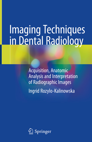
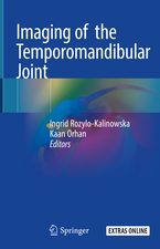
Imaging Techniques in Dental Radiology: Acquisition, Anatomic Analysis and Interpretation of Radiographic Images
Autor Ingrid Rozylo-Kalinowskaen Limba Engleză Hardback – aug 2020
| Toate formatele și edițiile | Preț | Express |
|---|---|---|
| Paperback (1) | 638.84 lei 38-44 zile | |
| Springer International Publishing – 2 aug 2021 | 638.84 lei 38-44 zile | |
| Hardback (1) | 894.62 lei 38-44 zile | |
| Springer International Publishing – aug 2020 | 894.62 lei 38-44 zile |
Preț: 894.62 lei
Preț vechi: 941.70 lei
-5% Nou
Puncte Express: 1342
Preț estimativ în valută:
171.21€ • 177.39$ • 142.88£
171.21€ • 177.39$ • 142.88£
Carte tipărită la comandă
Livrare economică 18-24 martie
Preluare comenzi: 021 569.72.76
Specificații
ISBN-13: 9783030413712
ISBN-10: 3030413713
Pagini: 188
Ilustrații: XIX, 188 p. 194 illus., 158 illus. in color.
Dimensiuni: 155 x 235 mm
Ediția:1st ed. 2020
Editura: Springer International Publishing
Colecția Springer
Locul publicării:Cham, Switzerland
ISBN-10: 3030413713
Pagini: 188
Ilustrații: XIX, 188 p. 194 illus., 158 illus. in color.
Dimensiuni: 155 x 235 mm
Ediția:1st ed. 2020
Editura: Springer International Publishing
Colecția Springer
Locul publicării:Cham, Switzerland
Cuprins
Introduction to Dental Radiography.- Materials and Preparation for Dental X-rays.- Intraoral Radiography in Dentistry.- Panoramic Radiography in Dentistry.- Cephalometric Radiograph in Dentistry.- Dental Cone-Beam Computed Tomography (CBCT).- Technical Errors and Artefacts in Dental Radiography.- Normal Anatomical Landmarks in Dental X-rays and CBCT Studies.- Analysis of Dental Radiographs and CBCT Studies.- Legal Provisions for Ionizing Radiation in Dentistry.- Safety Precautions for Dental Patient and Dental Staff Using X-ray.
Recenzii
“This is an excellent, compact book that is worth every penny and from which I am sure anyone involved with dental imaging, no matter how experienced, will learn something new.” (Louise Burton, RAD Magazine, March, 2021)
Notă biografică
Ingrid Różyło-Kalinowska, MD, PhD, DSc, is a specialist in radiology and diagnostic imaging who is currently Full Professor and Head of the Department of Dentomaxillofacial Radiology at the Medical University of Lublin in Poland. Her didactic work includes dental radiography and radiology, maxillofacial radiology, medical radiology, and diagnostic imaging for dentists, medical radiologists, and dental students. Dr. Różyło-Kalinowska is President of the European Academy of DentoMaxilloFacial Radiology (EADMFR), Regional Director of the International Association of DentoMaxilloFacial Radiology (IADMFR) in Europe, Vice-President of the Polish Dental Association, and Chair of the Section of DentoMaxillofacial Radiology of the Polish Medical Radiological Society. She will host the 18th European Congress of Dentomaxillofacial Radiology in Lublin in 2022. Dr. Różyło-Kalinowska belongs to the editorial boards of several scientific journals and is Editor-in-Chief of the “Journal ofStomatology”, official journal of the Polish Dental Association. To date, she has authored more than 200 full papers and co-authored five textbooks. She has also translated eight medical textbooks.
Textul de pe ultima copertă
This book is an up-to-date guide to the performance and interpretation of imaging studies in dental radiology. After opening discussion of the choice of X-ray equipment and materials, intraoral radiography, panoramic radiography, cephalometric radiology, and cone-beam computed tomography are discussed in turn. With the aid of many illustrated examples, patient preparation and positioning are thoroughly described for each modality. Common technical errors and artifacts are identified and the means of avoiding them, explained. The aim is to equip the reader with all the information required in order to perform imaging effectively and safely. The normal radiographic anatomy and landmarks are then discussed, prior to thorough coverage of frequent dentomaxillofacial lesions. Accompanying images display the characteristic features of each lesion. Further topics to be addressed are safety precautions for patients and staff. The book will be an ideal aid for all dental practitioners and will also be of value for dental students.
Caracteristici
Covers conventional imaging techniques and CBCT Explains how to avoid technical errors and artifacts Helps to identify frequent dentomaxillofacial lesions Includes numerous helpful illustrations
