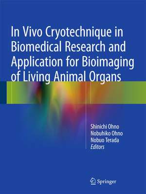
In Vivo Cryotechnique in Biomedical Research and Application for Bioimaging of Living Animal Organs
Editat de Shinichi Ohno, Nobuhiko Ohno, Nobuo Teradaen Limba Engleză Hardback – 6 dec 2015
| Toate formatele și edițiile | Preț | Express |
|---|---|---|
| Paperback (1) | 1030.83 lei 38-44 zile | |
| Springer – 28 mar 2019 | 1030.83 lei 38-44 zile | |
| Hardback (1) | 1126.45 lei 3-5 săpt. | |
| Springer – 6 dec 2015 | 1126.45 lei 3-5 săpt. |
Preț: 1126.45 lei
Preț vechi: 1185.73 lei
-5% Nou
Puncte Express: 1690
Preț estimativ în valută:
215.61€ • 234.28$ • 181.23£
215.61€ • 234.28$ • 181.23£
Carte disponibilă
Livrare economică 31 martie-14 aprilie
Preluare comenzi: 021 569.72.76
Specificații
ISBN-13: 9784431557227
ISBN-10: 4431557229
Pagini: 300
Ilustrații: XI, 300 p. 114 illus., 99 illus. in color.
Dimensiuni: 210 x 279 x 20 mm
Greutate: 1.22 kg
Ediția:1st ed. 2016
Editura: Springer
Colecția Springer
Locul publicării:Tokyo, Japan
ISBN-10: 4431557229
Pagini: 300
Ilustrații: XI, 300 p. 114 illus., 99 illus. in color.
Dimensiuni: 210 x 279 x 20 mm
Greutate: 1.22 kg
Ediția:1st ed. 2016
Editura: Springer
Colecția Springer
Locul publicării:Tokyo, Japan
Public țintă
ResearchCuprins
Preface.- Part. Overview.- Chapter 1. Introduction.- Chapter 2. Biomedical significance and development of “IVCT”.- Chapter 3. How to perform ‘IVCT’.- Chapter 4. Technical merits with ‘IVCT’.- Part Ⅱ. Application of ‘IVCT’ to various mouse organs.- Chapter 5. Histochemical Analyses of Living Mouse Liver under Different Hemodynamic Conditions.- Chapter 6. Three-dimensional reconstruction of liver tissues with confocal laser scanning microscopy.- Chapter 7. Application of “in vivo cryotechnique” to immunohistochemical detection of hypoxia in mouse liver tissues treated with pimonidazole.- Chapter 8. Immunohistochemical detection of soluble immunoglobulins in small intestines.- Chapter 9. Detection of MAPK signal transduction proteins in an ischemia/reperfusion model of small intestines.- Chapter 10. Histological Study and LYVE-1 Immunolocalization of Mesenteric Lymph Nodes.- Chapter 11. Distribution of immunoglobulin-producing cells in immunized mouse spleens.- Chapter12. Alteration of erythrocyte shapes in various organs of living mice or under blood flow conditions.- Chapter 13. Dynamic ultrastructure of smooth muscle cells in dystrophin-deficient mdx or normal scn mice.- Chapter 14. Dynamic Bioimaging of Serum Proteins in Beating Mouse Hearts.- Chapter 15. Histochemical analyses and quantum dot imaging of microvascular blood flow with pulmonary edema.- Chapter 16. Dynamic ultrastructure of pulmonary alveoli of living mice under respiratory conditions.- Chapter 17. Immunohistochemical analysis of various serum proteins in living mouse thymus.- Chapter 18. Immuno- and enzyme-histochemistry of HRP for demonstration of blood vessel permeability in mouse thymic tissues.- Chapter 19. Immunolocalization of serum proteins in living mouse glomeruli under various hemodynamic conditions.- Chapter 20. Immunohistochemical Analyses on Albumin and IgG in Acute Hypertensive Mouse Kidneys.- Chapter 21. Application of novel "in vivo cryotechnique" in living animal kidneys.- Chapter 22. Application of periodic acid-Schiff fluorescence emission for imunohistochemistry of living mouse renal glomeruli.- Chapter 23. Immunohistochemical analyses on serum proteins in nephrons of protein-overload mice.- Chapter 24. Immunohistochemical analyses of serum proteins in puromycin aminonucleoside nephropathy of living rat kidneys.- Chapter 25. Immunolocalization of phospho-Arg-directed protein kinase-substrate in hypoxic kidneys.- Chapter 26. Dynamic ultrastructures of renal glomeruli in living mice under various hemodynamic conditions.- Chapter 27. Function of dynamin-2 in formation of discoid vesicles in urinary bladder umbrella cells.- Chapter 28. Involvement of follicular basement membrane and vascular endothelium in blood-follicle barrier formation of mice.- Chapter 29. Permselectivity of blood-follicle barriers in mouse polycystic ovary model.- Chapter 30. Application of “in vivo cryotechnique” to immunohistochemical study of serum albumin in normal and cadmium-treated mouse testis organs.- Chapter 31. Immunohistochemical detection of angiotensin II receptors in mouse cerebellum.- Chapter 32. Application of “in vivo cryotechnique” to immunohistochemical analyses for effects of anoxia on serum immunoglobulin and albumin leakage through blood–brain barrier in mouse cerebellum.- Chapter 33. Extracellular Space in Central Nervous System.- Chapter 34. Overview on recent applications of in vivo cryotechnique in neurosciences.- Chapter 35. Application of “in vivo cryotechnique” to immunohistochemical detection of phosphorylated rhodopsin in light-exposed retina of living mouse.- Chapter 36. Application of “in vivo cryotechnique” to immunohistochemical detection of glutamate in mouse retina inner segment of photoreceptors.- Chapter 37. Application of “in vivo cryotechnique” to immunohistochemical study of mouse sciatic nerves under various stretching conditions.- Part Ⅲ. Further preparation after ‘IVCT’.- Chapter 38. Freeze-substitution (FS) fixation method.- Part IV. Comments on advantages of ‘IVCT’.- Chapter 39. Cryotechniques and freeze-substitution for immunostaining of intranuclear antigens and fluorescence in situ hybridization.- Chapter 40. Replica immunoelectron microscopy for caveolin in living smooth muscle cells.- Chapter 41. Application of “in vivo cryotechnique” to detection of injected fluorescence-conjugated IgG in living mouse organs.- Chapter 42. Application of “in vivo cryotechnique” to visualization of microvascular blood flow in mouse kidney by quantum dot injection.- Part V . Direct detection of molecules by Raman microscopy.- Chapter 43. Application of “in vivo cryotechnique” to Raman microscopy of freeze-dried mouse eyeball-slice.- Chapter 44. Application of ‘‘in vivo cryotechnique” to detection of erythrocyte oxygen saturation in mouse blood vessels with confocal Raman cryomicroscopy.- Part VI . Future development for human pathology.- Chapter 45. Imaging of thrombosis and microcirculation in mouse lungs of initial melanoma metastasis.- Chapter 46. Differential distribution of blood-derived proteins in xenografted human adenocarcinoma tissues.- Part VII . New cryobiopsy invention.- Chapter 47. Morphological and Histochemical Analysis of Living Mouse Livers by New ‘Cryobiopsy’ Technique.- Chapter 48. Recent Development of In Vivo Cryotechnique to Cryobiopsy for Living Animals.- Part VIII . Application of cryobiopsy for living animal tissues.- Chapter 49. Significance of cryobiopsy for morphological and immunohistochemical analyses of xenografted human lung cancer tissues and functional blood vessels.- Chapter 50. Immunolocalization of serum proteins in xenografted mouse model of human tumor cells.- Chapter 51. Histochemical approach of cryobiopsy for glycogen distribution in living mouse livers under fasting and local circulation loss conditions.- Part IX . New bioimaging of living animal tissues.- Chapter 52. Bioimaging of fluorescence-labeled mitochondria in subcutaneously grafted murine melanoma cells by the “in vivo cryotechnique” .- Chapter 53. Application of “in vivo cryotechnique” to visualization of ATP with luciferin-luciferase reaction in mouse skeletal muscles.- Chapter 54. Concluding remarks.
Notă biografică
Shinichi Ohno, M.D., Ph.D.
Professor
Department of Anatomy and Molecular Histology, Interdisciplinary Graduate School of Medicine and Engineering, University of Yamanashi
Nobuhiko Ohno, M.D., Ph.D.
Associate Professor
Department of Anatomy and Molecular Histology, Interdisciplinary Graduate School of Medicine and Engineering, University of Yamanashi
Nobuo Terada, M.D., Ph.D.
Professor
Division of Health Science, Shinshu University Graduate School of Medicine
Professor
Department of Anatomy and Molecular Histology, Interdisciplinary Graduate School of Medicine and Engineering, University of Yamanashi
Nobuhiko Ohno, M.D., Ph.D.
Associate Professor
Department of Anatomy and Molecular Histology, Interdisciplinary Graduate School of Medicine and Engineering, University of Yamanashi
Nobuo Terada, M.D., Ph.D.
Professor
Division of Health Science, Shinshu University Graduate School of Medicine
Textul de pe ultima copertă
This book focuses on actual morphofunctional findings of cells and tissues in living animal organs. Medical and biological scientists need to know the real in vivo morphology and immunolocalization of the molecular components in living animal organs. Recently, the live imaging of cells and tissues of animals with fluorescence-labeled proteins by gene manipulation has become more and more popular in biological fields. Current research, meanwhile, has revealed that immunohistochemical or morphological studies exclusively depend on living animal organs. The cryotechnique is one of the most useful tools for immunohistochemistry and bioimaging of animal organs. This book describes the epoch-making cryotechnique originally developed by the editors. The book also makes the management of living animal morphology more accessible not only for biomedical researchers but also for clinical doctors, providing a valuable resource work on the current perspectives of in vivo morphology.
Caracteristici
Presents applications of a unique cryotechnique, in vivo cryotechnique
Is written for both biomedical researchers and clinical doctors interested in morphology
Covers the latest trends in live imaging, immunohistochemistry, gene manipulation and new microscopy analysis
Is written for both biomedical researchers and clinical doctors interested in morphology
Covers the latest trends in live imaging, immunohistochemistry, gene manipulation and new microscopy analysis
