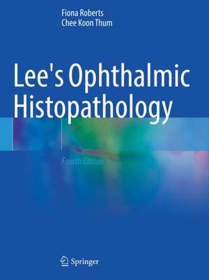
Lee's Ophthalmic Histopathology
Autor Fiona Roberts, Chee Koon Thumen Limba Engleză Paperback – 16 aug 2022
Well-illustrated and practically-oriented, this book has retained its general layout and style. The visual image remains key to explaining the pathological processes and this is facilitated by full color photography throughout the text. It helps both the pathologist and ophthalmologist understand the process that a specimen must go through prior to producing a report and how these various techniqueshelp to refine the diagnosis.
The fourth edition of Lee’s Ophthalmic Histopathology is an invaluable reference source for ophthalmic pathologists, general pathologists and ophthalmologists.
| Toate formatele și edițiile | Preț | Express |
|---|---|---|
| Paperback (2) | 876.19 lei 6-8 săpt. | |
| Springer International Publishing – 16 aug 2022 | 876.19 lei 6-8 săpt. | |
| SPRINGER LONDON – oct 2016 | 1191.42 lei 38-45 zile | |
| Hardback (1) | 1210.62 lei 3-5 săpt. | |
| Springer International Publishing – 15 aug 2021 | 1210.62 lei 3-5 săpt. |
Preț: 876.19 lei
Preț vechi: 922.31 lei
-5% Nou
Puncte Express: 1314
Preț estimativ în valută:
167.65€ • 175.68$ • 138.60£
167.65€ • 175.68$ • 138.60£
Carte tipărită la comandă
Livrare economică 11-25 aprilie
Preluare comenzi: 021 569.72.76
Specificații
ISBN-13: 9783030765279
ISBN-10: 303076527X
Pagini: 499
Ilustrații: XVII, 499 p. 750 illus., 728 illus. in color.
Dimensiuni: 210 x 279 mm
Greutate: 1.27 kg
Ediția:4th ed. 2021
Editura: Springer International Publishing
Colecția Springer
Locul publicării:Cham, Switzerland
ISBN-10: 303076527X
Pagini: 499
Ilustrații: XVII, 499 p. 750 illus., 728 illus. in color.
Dimensiuni: 210 x 279 mm
Greutate: 1.27 kg
Ediția:4th ed. 2021
Editura: Springer International Publishing
Colecția Springer
Locul publicării:Cham, Switzerland
Cuprins
Examination of the Globe: Technical Aspects.- The Traumatised Eye.- “Absolute Glaucoma”.- Retinal Vascular Disease.- Intraocular Tumours.- Ocular Inflammation.- Treatment of Retinal Detachment.- The Malformed Eye.- Autopsy Eye: the Eye in Systemic Disease.- Biopsy of the Eyelid, the Lacrimal Sac, and the Temporal Artery.- The Conjunctival Biopsy.- The Orbit: Biopsy, Excision Biopsy, and Exenteration Specimens.- The Corneal Disc.- Lens.
Notă biografică
Dr Fiona Roberts is a consultant pathologist with a special interest in Ophthalmic Pathology. During her training in pathology she undertook a period of research in ophthalmic pathology in Chicago following which she further trained in ophthalmic pathology in Glasgow with Professor Willliam Lee (the original author of the book). She has previous research interests in uveal and conjunctival melanoma and still undertakes research in toxoplasmosis (the subject of her research in Chicago). Her daily work involves reporting eye pathology specimens for the West of Scotland with a wider referral practice. She is also involved in teaching in the undergraduate medical and nursing curriculum and with trainee ophthalmologists and pathologists. She examines in pathology for the Royal College of Ophthalmologist.
She is an active member of the ophthalmic pathology community. She is a longstanding member of the British association of Ophthalmic Pathology and has completed terms as secretary and president (2013-2016) of the society. She is also an elected member of the European Ophthalmic Pathology Society since 2002 and is currently secretary and treasurer of this society.
She has been involved in publishing undergraduate medical text books including Pathology Illustrated (Editor) and Muir’s textbook of pathology (Chapter Author). She has also contributed to several ophthalmic pathology texts (in addition to Lee’s Ophthalmic Histopathology) including Eye Pathology, An illustrated guide (Chapter Author) and The Eye, Basic science in practice (Chapter Author). Other texts to which she has contributed include Adams and Graham’s Introduction to Neuropathology (Chapter Author) and Krugman’s Infectious Disease of Children (Chapter Author). She has also published over 80 original publications in journals.
Dr Chee Koon Thum is a consultant pathologist with a special interest in Ophthalmic Pathology and Molecular Pathology. He is currently working in both Royal Infirmary of Edinburgh (RIE) and Queen Elizabeth University Hospital (QEUH) in Glasgow, Scotland. He was originally trained in clinical ophthalmology and has been a member of the Royal College of Ophthalmologists and the Royal College of Surgeons (Edinburgh) for more than a decade. He continued to utilise his clinical knowledge and experience in his training of ophthalmic pathology with Dr Fiona Roberts (co-editor). He reports the eye pathology specimens in Edinburgh and Glasgow. Together with Dr Fiona Roberts, they deliver the ophthalmic pathology referral service for the rest of Scotland and Northern Ireland. He provides teaching for ophthalmology and pathology trainees. He is an active member of the British Association of Ophthalmic Pathology and has organised the annual meetings for the association. He is also the co-editor of the 3rd edition of Lee’s Ophthalmic Histopathology.
Textul de pe ultima copertă
Completely revised and updated, this book now outlines changing patterns in eye trauma, corneal surgery and glaucoma; developments in molecular genetics in uveal melanoma; and diagnostic techniques for intraocular lymphoma. Information on new treatments for vascular disease in diabetic eye disease and wet age related macular degeneration is now included and the chapter on the orbit has many new additions. Recently identified causes of inflammation in the eye including drug reactions are described, as are the effects of topical therapies in basal cell carcinoma.
Well-illustrated and practically-oriented, this book has retained its general layout and style. The visual image remains key to explaining the pathological processes and this is facilitated by full color photography throughout the text. It helps both the pathologist and ophthalmologist understand the process that a specimen must go through prior to producing a report and how these various techniques help to refine the diagnosis.
The fourth edition of Lee’s Ophthalmic Histopathology is an invaluable reference source for ophthalmic pathologists, general pathologists and ophthalmologists.
Caracteristici
Retains the popular, original specimen-based chapter layout Extensive colour illustrations of macro specimens and microspecimens Includes illustrations of immunohistochemistry, molecular analyses and electron microscopy
Recenzii
“This is a very comprehensive atlas and textbook on the macroscopic and microscopic histopathology of the embryonic, childhood, adolescent and adult eyes. … I recommend this book for all ophthalmology residents, fellows, and experts of eye diseases. Internists and pathologists will benefit from the highest quality full color details presented here. I have no reservations in recommending this book.” (Joseph J. Grenier, Amazon.com, September, 2015)
The approach of the book is unconventional; chapters reflect the categories of specimens sent to pathologists, rather than the conventional anatomical approach that only really works if one already has Ophthalmic Pathology experience. [T]his is unequivocally the "textbook of choice" for the trainee ophthalmic pathologist and general histopathologist. It is also highly recommended to trainee and senior ophthalmologists, and as a reference book to ophthalmic professionals and researchers. (MA Parsons, Eye News, 2004;10(4):84)
The ‘hands-on’ structure should make this book a mandatory item in the laboratory. (Holger Baatz, Ophthalmologica 2003;217(5):5)
[T]he intention of the author is to provide general pathologists with a concise but nevertheless complete bench-book and guide them through the intricacies of eye dissection, appropriate sampling, and microscopic examination. New data from literature have been included. The author's goals have been fully
met. The coverage is systematic and the writing exceptionally clear. This is an ideal book for practicing general pathologists who only occasionally see eye specimens and usually need considerable guidance and support while examining ophthalmic biopsies or surgically resected bulbi and other surgical ophthalmic specimens. As a general pathologist, I have consulted this book and found it to be a valuable source of information. Even though there are several excellent textbooks of ophthalmic pathology, this one would be definitely my first choice.(Doody’s Book Review Service™, 5 stars, Ivan Damjanov)
