Light and Specular Microscopy of the Cornea
Autor Katerina Jirsovaen Limba Engleză Paperback – 5 iun 2019
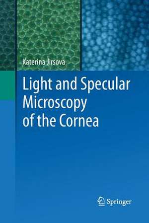
The atlas of the Light and Specular Microscopy of the Cornea, particularly of the corneal endothelium presents photographs of healthy and pathological corneas, as well as corneas prepared for grafting. Photographs are taken from donor or patient’s corneas. The first part section of the atlas shows healthy corneas and its particular layers: the epithelium (superficial and basal cells, subepithelial nerve plexus), stroma and keratocytes, and the endothelium. Blood vessels or palisades of Vogt in limbus are shown as well. The second part section that shows corneas processed for grafting is focused focuses on the endothelial layer. Main causes of exclusion of corneas from grafting, such as the presence of dead cells, polymeghatism, pleomorphism, cornea guttata or stromal scars have been shown. The third part section of the atlas shows corneas before and after storage in tissue cultures or hypothermic conditions with the aim to assess its suitability of for tissue for grafting. The last final section contains photographs of pathological corneal explants
| Toate formatele și edițiile | Preț | Express |
|---|---|---|
| Paperback (1) | 1595.70 lei 6-8 săpt. | |
| Springer International Publishing – 5 iun 2019 | 1595.70 lei 6-8 săpt. | |
| Hardback (1) | 1602.62 lei 6-8 săpt. | |
| Springer International Publishing – 6 feb 2018 | 1602.62 lei 6-8 săpt. |
Preț: 1595.70 lei
Preț vechi: 1679.67 lei
-5% Nou
Puncte Express: 2394
Preț estimativ în valută:
305.38€ • 317.64$ • 252.11£
305.38€ • 317.64$ • 252.11£
Carte tipărită la comandă
Livrare economică 14-28 aprilie
Preluare comenzi: 021 569.72.76
Specificații
ISBN-13: 9783319840291
ISBN-10: 3319840290
Pagini: 217
Ilustrații: XVIII, 217 p. 245 illus., 186 illus. in color.
Dimensiuni: 155 x 235 mm
Greutate: 0.34 kg
Ediția:Softcover reprint of the original 1st ed. 2017
Editura: Springer International Publishing
Colecția Springer
Locul publicării:Cham, Switzerland
ISBN-10: 3319840290
Pagini: 217
Ilustrații: XVIII, 217 p. 245 illus., 186 illus. in color.
Dimensiuni: 155 x 235 mm
Greutate: 0.34 kg
Ediția:Softcover reprint of the original 1st ed. 2017
Editura: Springer International Publishing
Colecția Springer
Locul publicării:Cham, Switzerland
Cuprins
1 The Cornea, Anatomy and Function, Katerina Jirsova.- 2 Processing Corneas for Grafting, Katerina Jirsova, Patricia Dahl, Jesper Hjortdal.- 3 Corneal Storage, Hypothermia, and Organ Culture, Katerina Jirsova, Patricia Dahl, W. John Armitage.- 4 Various Approaches to the Microscopic Assessment of the Cornea, Visualization and Image Analysis of the Corneal Endothelium, Katerina Jirsova, Jameson Clover, Christopher G. Stoeger, Gilles Thuret.- 5 Light and Specular Microscopy Assessment of the Cornea for Grafting, Katerina Jirsova, Jameson Clover, Christopher G. Stoeger, W. John Armitage.- 6 Atlas of Light and Specular Microscopy of the Cornea, Katerina Jirsova
Notă biografică
Katerina Jirsova
Laboratory of the Biology and Pathology of the Eye
Institute of Inherited Metabolic Disorders
Charles University First Faculty of Medicine,
Ke Karlovu 2, 128 08, Prague 2, Czech Republic
katerina.jirsova@lf1.cuni.cz
Textul de pe ultima copertă
The atlas of the Light and Specular Microscopy of the Cornea, particularly of the corneal endothelium presents photographs of healthy and pathological corneas, as well as corneas prepared for grafting. Photographs are taken from donor or patient’s corneas. The first part section of the atlas shows healthy corneas and its particular layers: the epithelium (superficial and basal cells, subepithelial nerve plexus), stroma and keratocytes, and the endothelium. Blood vessels or palisades of Vogt in limbus are shown as well. The second part section that shows corneas processed for grafting is focused focuses on the endothelial layer. Main causes of exclusion of corneas from grafting, such as the presence of dead cells, polymeghatism, pleomorphism, cornea guttata or stromal scars have been shown. The third part section of the atlas shows corneas before and after storage in tissue cultures or hypothermic conditions with the aim to assess its suitability of for tissue for grafting. The last final section contains photographs of pathological corneal explants
Caracteristici
The first published atlas of the cornea in light and specular microscopy which helps to assess and decide upon the suitability of corneas for grafting
Illustrative photographs attached to an educative text to discern healthy and pathological corneas
Photographs include corneas stored in tissue culture, hypothermic conditions, as well as pathological explants which increases both the appeal and the interest of the atlas to a wide audience
Illustrative photographs attached to an educative text to discern healthy and pathological corneas
Photographs include corneas stored in tissue culture, hypothermic conditions, as well as pathological explants which increases both the appeal and the interest of the atlas to a wide audience
