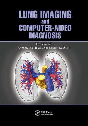
Lung Imaging and Computer Aided Diagnosis
Editat de Ayman El-Baz, Jasjit S. Surien Limba Engleză Paperback – 9 mar 2018
Lung Imaging and Computer Aided Diagnosis brings together researchers in pulmonary image analysis to present state-of-the-art image processing techniques for detecting and diagnosing lung cancer at an early stage. The book addresses variables and discrepancies in scans and proposes ways of evaluating small lung masses more consistently to allow for more accurate measurement of growth rates and analysis of shape and appearance of the detected lung nodules.
Dealing with all aspects of image analysis of the data, this book examines:
- Lung segmentation
- Nodule segmentation
- Vessels segmentation
- Airways segmentation
- Lung registration
- Detection of lung nodules
- Diagnosis of detected lung nodules
- Shape and appearance analysis of lung nodules
| Toate formatele și edițiile | Preț | Express |
|---|---|---|
| Paperback (1) | 552.60 lei 43-57 zile | |
| CRC Press – 9 mar 2018 | 552.60 lei 43-57 zile | |
| Hardback (1) | 1414.28 lei 43-57 zile | |
| CRC Press – 23 aug 2011 | 1414.28 lei 43-57 zile |
Preț: 552.60 lei
Preț vechi: 801.32 lei
-31% Nou
Puncte Express: 829
Preț estimativ în valută:
105.74€ • 110.68$ • 88.01£
105.74€ • 110.68$ • 88.01£
Carte tipărită la comandă
Livrare economică 31 martie-14 aprilie
Preluare comenzi: 021 569.72.76
Specificații
ISBN-13: 9781138072077
ISBN-10: 1138072079
Pagini: 496
Ilustrații: Approx. 425 to 475 equations.; 45 Tables, black and white; 30 Illustrations, color; 735 Illustrations, black and white
Dimensiuni: 178 x 254 x 32 mm
Greutate: 0.45 kg
Ediția:1
Editura: CRC Press
Colecția CRC Press
ISBN-10: 1138072079
Pagini: 496
Ilustrații: Approx. 425 to 475 equations.; 45 Tables, black and white; 30 Illustrations, color; 735 Illustrations, black and white
Dimensiuni: 178 x 254 x 32 mm
Greutate: 0.45 kg
Ediția:1
Editura: CRC Press
Colecția CRC Press
Cuprins
A Novel Three-Dimensional Framework for Automatic Lung Segmentation from Low- Dose Computed Tomography Images. Incremental Engineering of Lung Segmentation Systems. 3D MGRF-Based Appearance Modeling for Robust Segmentation of Pulmonary Nodules in 3D LDCT Chest Images. Ground-Glass Nodule Characterization in High-Resolution Computed Tomography Scans. Four-Dimensional Computed Tomography Lung Registration Methods. Pulmonary Kinematics via Registration of Serial Lung Images. Acquisition and Automated Analysis of Normal and Pathological Lungs in Small Animals Using Computed Microtomography. Airway Segmentation and Analysis from Computed Tomography. Pulmonary Vessel Segmentation for Multislice CT Data: Methods and Applications. A Novel Level Set-Based Computer-Aided Detection System for Automatic Detection of Lung Nodules in Low-Dose Chest Computed Tomography Scans. Model-Based Methods for Detection of Pulmonary Nodules. Concept and Practice of Genetic Algorithm Template Matching and Higher Order Local Autocorrelation Schemes in Automated Detection of Lung Nodules. Computer-Aided Detection of Lung Nodules in Chest Radiographs and Thoracic CT. Lung Nodule and Tumor Detection and Segmentation. Texture Classification in Pulmonary CT. Computer-Aided Assessment and Stenting of Tracheal Stenosis. Appearance Analysis for the Early Assessment of Detected Lung Nodules. Validation of a New Image-Based Approach for the Accurate Estimating of the Growth Rate of Detected Lung Nodules Using Real Computed Tomography Images and Elastic Phantoms Generated by State-of-the-Art Microfluidics Technology.Three-Dimensional Shape Analysis Using Spherical Harmonics for Early Assessment of Detected Lung Nodules. Index.
Notă biografică
Ayman El-Baz received BSc and MS degrees in electrical engineering from Mansoura University, Egypt, in 1997 and 2000, respectively, and a PhD degree in electrical engineering from University of Louisville, Kentucky. He joined the Bioengineering Department of the University of Louisville in August 2006. His current research is focused on developing new computer-assisted diagnosis systems for different diseases and brain disorders.
Jasjit S. Suri is an innovator, a scientist, a visionary, an industrialist, and an internationally known world leader in biomedical engineering. Dr. Suri has spent over 20 years in the field of biomedical engineering/devices and its management. He received his doctorate from the University of Washington, Seattle, and a business management sciences degree from Weatherhead, Case Western Reserve University, Cleveland, Ohio. Dr. Suri was awarded the President’s Gold Medal in 1980 and the Fellow of American Institute of Medical and Biological Engineering for his outstanding contributions.
Jasjit S. Suri is an innovator, a scientist, a visionary, an industrialist, and an internationally known world leader in biomedical engineering. Dr. Suri has spent over 20 years in the field of biomedical engineering/devices and its management. He received his doctorate from the University of Washington, Seattle, and a business management sciences degree from Weatherhead, Case Western Reserve University, Cleveland, Ohio. Dr. Suri was awarded the President’s Gold Medal in 1980 and the Fellow of American Institute of Medical and Biological Engineering for his outstanding contributions.
Descriere
There is an urgent need for new technology to diagnose small, malignant lung nodules early as well as large nodules located away from large diameter airways because current technology—namely, needle biopsy and bronchoscopy—fail to diagnose those cases. This book brings together researchers in pulmonary image analysis to discuss state-of-the-art image processing techniques for detecting and diagnosing lung cancer at an early stage. The book addresses variables and discrepancies in scans and proposes ways of evaluating small lung masses more consistently to allow for more accurate measurement of growth rates and analysis of shape and appearance of the detected lung nodules.
