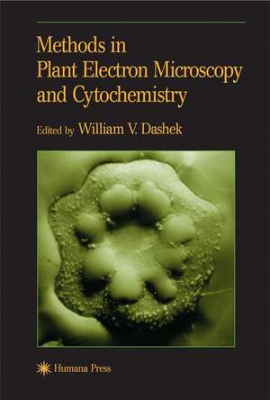
Methods in Plant Electron Microscopy and Cytochemistry
Editat de William V. Dasheken Limba Engleză Paperback – 29 iun 2000
| Toate formatele și edițiile | Preț | Express |
|---|---|---|
| Paperback (2) | 640.55 lei 43-57 zile | |
| Humana Press Inc. – 29 iun 2000 | 640.55 lei 43-57 zile | |
| Humana Press Inc. – 5 noi 2010 | 999.14 lei 43-57 zile |
Preț: 640.55 lei
Preț vechi: 753.60 lei
-15% Nou
Puncte Express: 961
Preț estimativ în valută:
122.57€ • 128.31$ • 101.42£
122.57€ • 128.31$ • 101.42£
Carte tipărită la comandă
Livrare economică 07-21 aprilie
Preluare comenzi: 021 569.72.76
Specificații
ISBN-13: 9780896038097
ISBN-10: 0896038092
Pagini: 300
Ilustrații: XI, 300 p. 81 illus., 7 illus. in color.
Dimensiuni: 152 x 229 x 17 mm
Greutate: 0.42 kg
Ediția:2000
Editura: Humana Press Inc.
Colecția Humana
Locul publicării:Totowa, NJ, United States
ISBN-10: 0896038092
Pagini: 300
Ilustrații: XI, 300 p. 81 illus., 7 illus. in color.
Dimensiuni: 152 x 229 x 17 mm
Greutate: 0.42 kg
Ediția:2000
Editura: Humana Press Inc.
Colecția Humana
Locul publicării:Totowa, NJ, United States
Public țintă
ResearchCuprins
1 Plant Cells and Tissues: Structure—Function Relationships.- 2 Methods for the Cytochemical/Histochemical Localization of Plant Cell/Tissue Chemicals.- 3 Methods in Light Microscope Radioautography.- 4 Some Fluorescence Microscopical Methods for Use with Algal, Fungal, and Plant Cells.- 5 Fluorescence Microscopy of Aniline Blue Stained Pistils.- 6 A Short Introduction to Immunocytochemistry and a Protocol for Immunovisualization of Proteins with Alkaline Phosphatase.- 7 The Fixation of Chemical Forms on Nitrocellulose Membranes.- 8 Dark-Field Microscopy and Its Application to Pollen Tube Culture.- 9 Computer-Assisted Microphotometry.- 10 Isolation and Characterization of Endoplasmic Reticulum from Mulberry Cortical Parenchyma Cells.- 11 Methods for the Identification of Isolated Plant Cell Organelles.- 12 Methods for the Detergent Release of Particle-Bound Plant Proteins.- 13 Scanning Electron Microscopy: Preparations for Diatoms.- 14 Methods for the Ultrastructural Analysis of Plant Cells and Tissues.- 15 Methods for Atomic Force and Scanning Tunneling Microscopies.- 16 Visualization of Golgi Apparatus by Zinc Iodide—Osmium Tetroxide (ZIO) Staining.- 17 Methods in Electron Microscope Autoradiography.- 18 Electron Microscopic Immunogold Localization.- 19 Microanalysis.- 20 The Use of Electron Microscopy in Molecular Biology.- 21 Summation.
Recenzii
"...contributing authors discuss novel, contemporary microscopic technologies needed for the effective study of plant cell biology in a user-friendly manner. The book provides conceptual information as well as protocols for immediate microscopic application...Following an introductory chapter on plant cell structure-function relationships, the author offers some recent developments regarding fixation, dehydration and embedding of plant cells and tissues for the light microscopic localization of certain of their chemical constituents...Separate chapters are devoted to light microscope radioautography, electron microscope autoradiography, fluorescence microscopy, dark-field microscopy, immunocytochemistry, electron microscopic immunogold localization and computer assisted microphotometry...The manual, excellently combining background theory with practical applications, can be useful to graduate and undergraduate students, postdoctorals, professors, as well as government and industrial scientists." - Acta Botanica Hungarica
"...very useful to researchers just beginning to delve into plant research. It is written for the most part in a very easy to understand way and is not filled with jargon. There are many tables that contain concise summaries of pertinent information about methods, structures, stains, etc....describes the basic structures found within plant cells, including the size ranges of the organelles. This information is very helpful to those of us who sometimes forget how big a ribosome should be. The included micrographs are nice that they show the reader what certain structures should look like on the TEM...useful addition to the library of a plant researcher." - Plant Science Bulletin
"Easy-to-use and up-to-date, it offers today's plant scientists a first class collection of readily reproducible light and electron microscopical methods that will prove the new standard for all working in the field." - Lavoisier Librairie
"This lab manual is certainly extremely valuable for research specialists from the plant area as well as indispensable for the graduate students starting their research career...this book will provide a valuable source for several years to come for advanced scientists working in the field of plant structures." -Cell Biology International
Textul de pe ultima copertă
Although there are many electron microscopy monographs concerned with animal cells, there are few devoted to plant cells and tissues. In Methods in Plant Electron Microscopy and Cytochemistry, hands-on experimentalists describe the cutting-edge microscopical methods needed for the effective study of plant cell biology today. These powerful techniques, all described in great detail to ensure successful experimental results, range from light microscope cytochemistry, autoradiography, and immunocytochemistry, to recent developments in fluorescence, confocal, and dark-field microscopies. Important advances in both conventional and scanning electron microscopies are also carefully developed, together with such state-of-the-art ancillary techniques as high-resolution autoradiography, immunoelectron microscopy, x-ray microanalysis, and electron systems imaging. Applications are for the most part demonstrated with higher plants, while some are applied to studies of fungi.
Easy-to-use and up-to-date, Methods in Plant Electron Microscopy and Cytochemistry offers todayís plant scientists a firstclass collection
of readily reproducible light and electron microscopical methods that will prove the new standard for all working in the field.
Easy-to-use and up-to-date, Methods in Plant Electron Microscopy and Cytochemistry offers todayís plant scientists a firstclass collection
of readily reproducible light and electron microscopical methods that will prove the new standard for all working in the field.
Caracteristici
Includes supplementary material: sn.pub/extras
