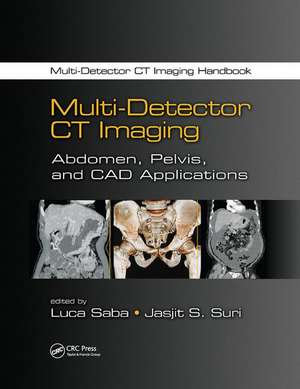
Multi-Detector CT Imaging: Abdomen, Pelvis, and CAD Applications
Editat de Luca Saba, Jasjit S. Surien Limba Engleză Paperback – 22 noi 2017
It is no wonder that with the critical role CT plays and the rapid innovations in computer technology that advances in the capabilities and complexity of CT imaging continue to evolve. While information about these developments may be scattered about in journals and other resources, this two-volume set provides an authoritative, up-to-date, and educational reference that covets the entire spectrum of CT.
| Toate formatele și edițiile | Preț | Express |
|---|---|---|
| Paperback (2) | 427.84 lei 6-8 săpt. | |
| CRC Press – 22 noi 2017 | 427.84 lei 6-8 săpt. | |
| CRC Press – 22 noi 2017 | 449.79 lei 6-8 săpt. | |
| Hardback (2) | 1128.78 lei 6-8 săpt. | |
| CRC Press – 21 oct 2013 | 1128.78 lei 6-8 săpt. | |
| CRC Press – 21 oct 2013 | 1241.06 lei 6-8 săpt. |
Preț: 427.84 lei
Preț vechi: 552.56 lei
-23% Nou
Puncte Express: 642
Preț estimativ în valută:
81.89€ • 85.17$ • 68.62£
81.89€ • 85.17$ • 68.62£
Carte tipărită la comandă
Livrare economică 13-27 martie
Preluare comenzi: 021 569.72.76
Specificații
ISBN-13: 9781138072527
ISBN-10: 1138072524
Pagini: 692
Ilustrații: 81 Tables, black and white; 723 Illustrations, black and white
Dimensiuni: 210 x 280 x 40 mm
Greutate: 0.45 kg
Ediția:1
Editura: CRC Press
Colecția CRC Press
ISBN-10: 1138072524
Pagini: 692
Ilustrații: 81 Tables, black and white; 723 Illustrations, black and white
Dimensiuni: 210 x 280 x 40 mm
Greutate: 0.45 kg
Ediția:1
Editura: CRC Press
Colecția CRC Press
Public țintă
Academic and Professional Practice & DevelopmentCuprins
Liver. Computed Tomography of the Gallbladder and Bile Ducts. Spleen. The Pancreas. The Adrenal Gland. Pathology of the Stomach and Small Bowel. Imaging of the Retroperitoneum. Abdominal Wall. Diseases of the Colon and Rectum. Posttraumatic and Postsurgical Abdomen. Kidney and Ureters. Bladder. Male Pelvis. Female Pelvis: Uterus, Ovaries, Fallopian Tubes, and Vagina. Degenerative and Traumatic Diseases of the Spine. Inflammatory Disease of the Spine. Imaging of Spinal Tumors with Emphasis on MDCT. Ilio-Sacral Pathology. Upper and Lower Extremities. Pathology of the Muscles and Soft Tissues. Computed Tomography Based Interventional Radiology in the Musculoskeletal System. Pediatric Application. Forensic Medicine. Dental Scan and Odontoiatric Applications. Dual Energy CT: Tissue Characterization. Computed Tomography Multispectral Imaging. Positron Emission Tomography–Computed Tomography: Concept and General Application. Modified Akaike Information Criterion for Selecting the Numbers of Mixture Components: An Application to Initial Lung Segmentation. CAD Computed Tomography Lung Application. PET/CT Nodule Segmentation and Diagnosis: A Survey. Index.
Descriere
Computed Tomography represents a widely used diagnostic imaging technique in the medical field. In the last 20 years. CT technology has dramatically improved. CT scanners can image human bodies with an exceptional spatial resolution by diagnosing with high sensitivity and specificity several kinds of pathologies. This book offers a complete and clear description of the CT technique and potentialities in diagnosing human pathologies. All of the organs are covered in detail.
Recenzii
"This book starts with a chapter on technical principles of CT that explains the basic physics in a lucid manner. There are chapters on contrast media and also on radiation dose that cover the necessary requirements of postgraduate examinations. In the system-wise chapters, this book is a good treatise on the imaging of those organs which have still a major role or exclusive role of CT, e.g., lungs, pancreas, and clinical conditions like trauma/emergency imaging. The book does mention MRI appearances and includes MR images where necessary, e.g., neuroimaging. The book has been printed on good quality paper and the binding is strong. ... The book will be a good addition on the radiologist’s bookshelf and will be useful for day-to-day reference especially for emergency and trauma CT reporting."
—Mandar Varadpande, The Society of Radiologists in Training
—Mandar Varadpande, The Society of Radiologists in Training
Notă biografică
Luca Saba, University of Cagliari, Italy. Jasjit S. Suri, Global Biomedical Technologies Inc, Roseville USA.
