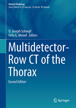
Multidetector-Row CT of the Thorax: Medical Radiology
Editat de U. Joseph Schoepf, Felix G. Meinelen Limba Engleză Hardback – 12 iul 2016
Din seria Medical Radiology
- 5%
 Preț: 1108.87 lei
 - 5%
 Preț: 349.24 lei
 - 5%
 Preț: 1308.02 lei
 - 5%
 Preț: 1308.74 lei
 - 5%
 Preț: 720.68 lei
 - 5%
 Preț: 717.20 lei
 - 5%
 Preț: 1626.03 lei
 - 5%
 Preț: 1618.70 lei
 - 5%
 Preț: 802.21 lei
 - 5%
 Preț: 1130.07 lei
 - 5%
 Preț: 1116.00 lei
 - 5%
 Preț: 1953.34 lei
 - 5%
 Preț: 783.04 lei
 - 5%
 Preț: 1105.61 lei
 - 5%
 Preț: 794.00 lei
 - 5%
 Preț: 1101.21 lei
 - 5%
 Preț: 821.19 lei
 - 5%
 Preț: 1420.29 lei
 - 5%
 Preț: 743.16 lei
 - 5%
 Preț: 906.63 lei
 - 5%
 Preț: 1313.75 lei
 - 5%
 Preț: 1858.30 lei
 - 5%
 Preț: 1306.73 lei
 - 5%
 Preț: 1113.11 lei
 - 5%
 Preț: 1462.37 lei
 - 5%
 Preț: 1301.44 lei
 - 5%
 Preț: 975.17 lei
 - 5%
 Preț: 1122.58 lei
 - 5%
 Preț: 1986.27 lei
 - 5%
 Preț: 1126.82 lei
 - 5%
 Preț: 718.46 lei
 - 5%
 Preț: 1450.84 lei
 - 5%
 Preț: 1298.14 lei
 - 5%
 Preț: 1110.32 lei
 - 5%
 Preț: 1184.42 lei
 - 5%
 Preț: 1113.99 lei
 - 5%
 Preț: 1435.85 lei
 - 5%
 Preț: 663.23 lei
 - 5%
 Preț: 1605.11 lei
 - 5%
 Preț: 731.07 lei
 - 5%
 Preț: 733.09 lei
 - 5%
 Preț: 1124.07 lei
 - 5%
 Preț: 383.93 lei
 - 5%
 Preț: 1106.69 lei
 - 5%
 Preț: 982.50 lei
 - 5%
 Preț: 1317.17 lei
 - 5%
 Preț: 1437.67 lei
 - 5%
 Preț: 1307.85 lei
 - 5%
 Preț: 1950.60 lei
Preț: 1355.82 lei
Preț vechi: 1427.18 lei
-5% Nou
Puncte Express: 2034
Preț estimativ în valută:
259.47€ • 269.89$ • 214.21£
259.47€ • 269.89$ • 214.21£
Carte tipărită la comandă
Livrare economică 11-17 aprilie
Preluare comenzi: 021 569.72.76
Specificații
ISBN-13: 9783319303536
ISBN-10: 3319303538
Pagini: 405
Ilustrații: XVIII, 597 p. 420 illus., 160 illus. in color.
Dimensiuni: 178 x 254 x 38 mm
Greutate: 1.68 kg
Ediția:2nd ed. 2016
Editura: Springer International Publishing
Colecția Springer
Seriile Medical Radiology, Diagnostic Imaging
Locul publicării:Cham, Switzerland
Cuprins
Part I: MDCT – Technical Background, Radiation Protection: Technical Bases of MDCT.- Radiation Exposure in Thoracic CT.- Strategies for Dose Reduction and Improvement of Image Quality in Chest CT.- Contrast Medium Injection Techniques.- Acquisition Protocols for Thoracic CT.- Part II: Airways / Diffuse Lung Disease: CT of the Airways.- CT in COPD / Pulmonary Emphysema.- CT Imaging of Interstitial Lung Diseases.- Pulmonary infections: Imaging with CT.- CT Evaluation of ARDS.- Part III: Lung Nodules / Lung Cancer: CT Screening for Lung Cancer: Evidence and Recommendations.- CT Screening for Lung Cancer: Current Controversies.- CT Characterization of Lung Nodules.- Management of lung nodules detected incidentally and at screening.- Staging of Lung Cancer with CT.- CT Imaging of the Mediastinum.- PET CT of the Thorax.- Part IV: Cardiovascular Applications: CT of Pulmonary Thromboembolic Disease.- Dual-energy CT of the Thorax.- MDCT of the Thoracic Aorta.- CT Imaging of Ischemic Heart Disease.- Comprehensive CT Imaging in acute chest pain.- CT Imaging of the Heart-Lung axis.- Anomalies and malformations of the pulmonary circulation: Evaluation with CT.- Part V: Data Management: Workflow Design for CT of the Thorax.- 2D and 3D Visualization of Thoracic MDCT Data.- Computer-Aided Diagnosis in Chest CT.- Part VI: Miscellaneous: Pediatric CT of the Chest.- CT of the Chest Wall.- CT in Chest Trauma.- CT Guided Interventions in the Thorax.- Thoracic CT: Medicolegal Aspects.- Future Developments in Chest CT.
Notă biografică
Joe Schoepf is a Professor with appointments in Radiology, Medicine, and Pediatrics at the Medical University of South Carolina (MUSC) in Charleston, SC. At MUSC Dr. Schoepf serves as director of the Division of Cardiovascular Imaging, of CT Research and Development, and as Cardiovascular Imaging Director of the University Designated Center for Biomedical Imaging.
Dr. Schoepf, a native of Austria, graduated from the medical school of Ludwig Maximilians University in Munich, Germany, in 1996. After his residency in Diagnostic Radiology at Klinikum Grosshadern, Munich, Germany, he assumed a position at Brigham and Women’s Hospital, Harvard Medical School, in Boston, MA, which he held from 2001-2004. His main clinical and scientific interest is non-invasive cardiovascular and thoracic imaging, especially the use of advanced CT and MRI techniques for diagnosing disorders of the heart and lung.
Felix G. Meinel is a clinical fellow and researcher in the Departmentof Clinical Radiology at Ludwig-Maximilians-University Hospital in Munich, Germany. Dr. Meinel received his medical degree from Ludwig-Maximilians-University in Munich and trained in Radiology at the same institution. He was a visiting instructor in the Division of Cardiovascular Imaging at the Medical University of South Carolina (Charleston, SC) from 2013 to 2014 before returning to Munich to complete his clinical training. His main research interests include thoracic and cardiovascular CT imaging – both the advancement of CT technique and how CT imaging can be used to gain novel insights into disease.
Textul de pe ultima copertă
Since the first edition of this book was published in 2004, computed tomography has seen groundbreaking technical innovations that have transformed the field of thoracic imaging and opened novel possibilities for the detection of thoracic pathologies. This book highlights cutting-edge thoracic applications of CT imaging in the context of these technical innovations and discusses the latest opportunities, with critical appraisal of challenges and controversies. All topics are covered by renowned international experts. Chapters from the original edition have been thoroughly updated to reflect the state of the art in technology and scientific evidence, and new contributions included on recent developments such as dual-energy CT and CT imaging in patients with acute chest pain. The book is abundantly illustrated with high-quality images and illustrations.
Caracteristici
Provides a comprehensive overview of thoracic applications for CT Discusses clinical applications in the context of the latest technical developments Written by distinguished experts in the respective fields
