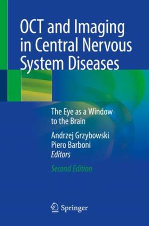
OCT and Imaging in Central Nervous System Diseases: The Eye as a Window to the Brain
Editat de Andrzej Grzybowski, Piero Barbonien Limba Engleză Paperback – 12 mar 2021
Similar to the first edition, this book is an excellent and richly illustrated reference for diagnosis of many retinaldiseases and monitoring of surgical and medical treatment. OCT allows to study vision from of the retina to the optic tracts. Retinal axons in the retinal nerve fiber layer (RNFL) are non-myelinated until they penetrate the lamina cribrosa. Hence, the RNFL is an ideal structure for visualization of any process of neurodegeneration, neuroprotection, or regeneration. By documenting the ability of OCT to provide key information on CNS diseases, this book illustrates convincingly that the eye is indeed the “window to the brain”.
| Toate formatele și edițiile | Preț | Express |
|---|---|---|
| Paperback (1) | 737.77 lei 38-44 zile | |
| Springer International Publishing – 12 mar 2021 | 737.77 lei 38-44 zile | |
| Hardback (1) | 1006.06 lei 38-44 zile | |
| Springer International Publishing – 2 ian 2020 | 1006.06 lei 38-44 zile |
Preț: 737.77 lei
Preț vechi: 776.60 lei
-5% Nou
Puncte Express: 1107
Preț estimativ în valută:
141.17€ • 147.41$ • 116.57£
141.17€ • 147.41$ • 116.57£
Carte tipărită la comandă
Livrare economică 11-17 aprilie
Preluare comenzi: 021 569.72.76
Specificații
ISBN-13: 9783030262716
ISBN-10: 3030262715
Pagini: 561
Ilustrații: X, 561 p. 159 illus., 141 illus. in color.
Dimensiuni: 155 x 235 mm
Ediția:2nd ed. 2020
Editura: Springer International Publishing
Colecția Springer
Locul publicării:Cham, Switzerland
ISBN-10: 3030262715
Pagini: 561
Ilustrații: X, 561 p. 159 illus., 141 illus. in color.
Dimensiuni: 155 x 235 mm
Ediția:2nd ed. 2020
Editura: Springer International Publishing
Colecția Springer
Locul publicării:Cham, Switzerland
Cuprins
Introduction: Retina Imaging – Past and Present.- OCT Technique – Past, Present and Future.- Optical Coherence Tomography and Optic Nerve Edema.- OCT and Compressive Optic Neuropathy.- Optical Coherence Tomography (OCT) and Multiple Sclerosis (MS).- OCT and Parkinson’s Disease.- Optical Coherence Tomography in Alzheimer’s Disease.- Friedreich’s Ataxia and More: Optical Coherence Tomography Findings in Rare Neurological Syndromes.- Other Neurological Disorders: Migraine, Neurosarcoidosis, Schizophrenia, Obstructive Sleep Apnea-Hypopnea Syndrome (OSAHS).- Hereditary Optic Neuropathies.- Trans Neuronal Retrograde Degeneration to OCT in Central Nervous System Diseases.- OCT in Toxic and Nutritional Optic Neuropathies.- Animal Models in Neuro Ophthalmology.- Optical Coherence Tomography (OCT) in Glaucoma.- OCT in Amblyopia.- Conclusion: The Exciting Future of OCT Imaging of Retina.
Notă biografică
Andrzej Grzybowski, M.D., Ph.D., MBA, is a Professor of Ophthalmology and Chair of Department of Ophthalmology, University of Warmia and Mazury, Olsztyn, Poland; Head of Institute for Research in Ophthalmology, Foundation for Ophthalmology Development, Poznan, Poland.
He is active in international scientific societies including Euretina (Co-opted Board member 2016-2018), Retina Society, AAO (International Fellow; member of the Global ONE Advisory Board and Museum of Vision’s Program Committee), EVER (Board member and chair of cataract section), ESCRS (curator of ESCRS Archive), and ISRS (member of the ISRS International Council), ISBCS, International Council of Ophthalmology (programme coordinator for WCO in 2011-2018), and Cogan Society. He became lifelong member (chair LIV) of the European Academy of Ophthalmology and its Treasurer. He has been active contributor to major ophthalmic conferences worldwide, including AAO (Achievement Award 2017, International Scholar Award 2018), APAO (International Coordinator 2017, Achievement Award 2018), WCO (Programme Coordinator 2010-2018), Euretina, ESCRS, EVER, ISOPT, etc.
He has been active editor, editor in chief and author of more than 450 peer-reviewed international publications (total IF higher than 1000), and over 50 book’s chapters; reviewer for more than 20 journals. He is a member of editorial boards of American Journal of Ophthalmology (IF 4.795), Acta Ophthalmologica (IF 3.157), PLOS One (IF 2.806), Graefe’s Archive for Clinical and Experimental Ophthalmology (IF 2,349), Translational Vision Science & Technology (TVST), (IF 2.221), BMC Ophthalmology (IF 1.586), Clinics in Dermatology (IF 2.253), Journal of Clinical Medicine (IF 5.8); Frontiers in Neurology (IF 3.5), Neuro-Ophthalmology (IF 0.2), Saudi Journal of Ophthalmology, Asia Pacific Academy of Ophthalmology Journal, and editor in chief of Archives of History and Philosophy of Medicine and Historia Ophthalmologica Internationalis.
Piero Barboni, MD, graduated in Medicine and Surgery from the University of Bologna, Italy, in 1986 and went on to complete his Specialization in Ophthalmology at the same university in 1990. After spending almost two decades in private practice, he was appointed Professor of Neuro-ophthalmology in the Department of Neurological Science, Bologna University. Since 2012 Dr. Barboni has been a Consultant Ophthalmologist at the Scientific Institute San Raffaele, University of Milan. His memberships include the Association for Research in Vision and Ophthalmology (ARVO) and the North American Neuro-Ophthalmology Society (NANOS). Dr. Barboni has been sub-investigator in many clinical trials on good clinical practice in multiple sclerosis and is a participant in an international research project on Leber’s hereditary optic neuropathy. He is the author of more than 70 articles in peer-reviewed journals.
Textul de pe ultima copertă
The second edition of OCT and Imaging in Central Nervous System Diseases offers updated state-of-the-art advances using optical coherence tomography (OCT) regrading neuronal loss within the retina. Detailed information on the OCT imaging and interpretation is provided for the evaluation of disease progression in numerous neurodegenerative disorders and as a biological marker of neuroaxonal injury. Covering disorders like multiple sclerosis, Parkinson’s disease, Alzheimer’s disease, intracranial hypertension, Friedreich’s ataxia, schizophrenia, hereditary optic neuropathies, glaucoma, and amblyopia, readers will given insights into effects on the retina and the and optic nerve. Individual chapters are also devoted to OCT technique, new OCT technology in neuro-ophthalmology, OCT and pharmacological treatment, and the use of OCT in animal models.
Similar to the first edition, this book is an excellent and richly illustrated reference for diagnosis of many retinaldiseases and monitoring of surgical and medical treatment. OCT allows to study vision from of the retina to the optic tracts. Retinal axons in the retinal nerve fiber layer (RNFL) are non-myelinated until they penetrate the lamina cribrosa. Hence, the RNFL is an ideal structure for visualization of any process of neurodegeneration, neuroprotection, or regeneration. By documenting the ability of OCT to provide key information on CNS diseases, this book illustrates convincingly that the eye is indeed the “window to the brain”.
Similar to the first edition, this book is an excellent and richly illustrated reference for diagnosis of many retinaldiseases and monitoring of surgical and medical treatment. OCT allows to study vision from of the retina to the optic tracts. Retinal axons in the retinal nerve fiber layer (RNFL) are non-myelinated until they penetrate the lamina cribrosa. Hence, the RNFL is an ideal structure for visualization of any process of neurodegeneration, neuroprotection, or regeneration. By documenting the ability of OCT to provide key information on CNS diseases, this book illustrates convincingly that the eye is indeed the “window to the brain”.
Caracteristici
Broadens understanding of OCT imaging and interpretation in clinical practice Discusses the role of OCT in evaluating disease progression in neurodegenerative disorders Presents translational aspects from animal models to the clinical approach
