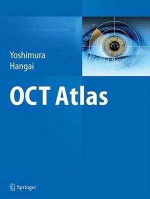
OCT Atlas
Autor Nagahisa Yoshimura, Masanori Hangaien Limba Engleză Paperback – 30 sep 2016
| Toate formatele și edițiile | Preț | Express |
|---|---|---|
| Paperback (1) | 813.79 lei 38-44 zile | |
| Springer Berlin, Heidelberg – 30 sep 2016 | 813.79 lei 38-44 zile | |
| Hardback (1) | 1160.09 lei 38-44 zile | |
| Springer Berlin, Heidelberg – 23 iun 2014 | 1160.09 lei 38-44 zile |
Preț: 813.79 lei
Preț vechi: 856.62 lei
-5% Nou
Puncte Express: 1221
Preț estimativ în valută:
155.74€ • 169.11$ • 130.82£
155.74€ • 169.11$ • 130.82£
Carte tipărită la comandă
Livrare economică 18-24 aprilie
Preluare comenzi: 021 569.72.76
Specificații
ISBN-13: 9783662513668
ISBN-10: 3662513668
Pagini: 364
Ilustrații: XIII, 351 p.
Dimensiuni: 210 x 279 x 26 mm
Greutate: 0.98 kg
Ediția:Softcover reprint of the original 1st ed. 2014
Editura: Springer Berlin, Heidelberg
Colecția Springer
Locul publicării:Berlin, Heidelberg, Germany
ISBN-10: 3662513668
Pagini: 364
Ilustrații: XIII, 351 p.
Dimensiuni: 210 x 279 x 26 mm
Greutate: 0.98 kg
Ediția:Softcover reprint of the original 1st ed. 2014
Editura: Springer Berlin, Heidelberg
Colecția Springer
Locul publicării:Berlin, Heidelberg, Germany
Cuprins
1 The basis of OCT interpretation.- 2 Vitreoretinal interface pathology.- 3 Diabetic retinopathy.- 4 Retinal vascular diseases.- 5 Central serous chorioretinopathy.- 6 Age-related macular degeneration.- 7 Retinal degeneration.- 8 Uveitis.- 9 Pathologic myopia and related diseases.- 10 Retinal detachment.- 11 Lesion morphology index based on OCT.- Service Part.
Notă biografică
Nagahisa Yoshimura, MD, PhD, Professor and Chairman, Department of Ophthalmology and Visual Scieces Kyoto University Graduate School of Medicine, Kyoto, Japan.
Masanori Hangai, MD, PhD, Professor and Chairman, Department of Ophthalmology, Saitama Medical University, Saitama, Japan.
Masanori Hangai, MD, PhD, Professor and Chairman, Department of Ophthalmology, Saitama Medical University, Saitama, Japan.
Caracteristici
Written by experts
Numerous color images
Numerous color images
Descriere
OCT provided a great advantage over other diagnostic modalities, as it could noninvasively provide tomographic images of the retina of a living eye. As a result, a number of new findings in retinal diseases were made using the time-domain OCT. OCT has now become an essential medical equipment OCT has now become an essential medical equipment in ophthalmic care and quality textbooks describing the functionality of OCT are very important in the education of young ophthalmologists and eye care personnel. In this book are chosen high quality OCT images of rather common diseases as well as images of several rare diseases.
