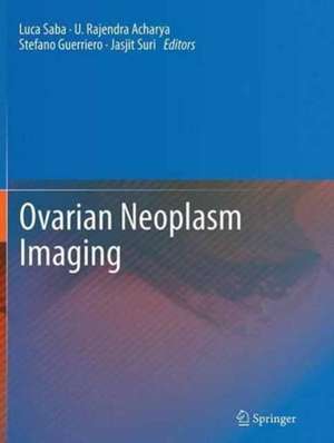
Ovarian Neoplasm Imaging
Editat de Luca Saba, U. Rajendra Acharya, Stefano Guerriero, Jasjit S. Surien Limba Engleză Paperback – 29 sep 2016
| Toate formatele și edițiile | Preț | Express |
|---|---|---|
| Paperback (1) | 1353.72 lei 38-44 zile | |
| Springer Us – 29 sep 2016 | 1353.72 lei 38-44 zile | |
| Hardback (1) | 1408.58 lei 38-44 zile | |
| Springer Us – 23 ian 2014 | 1408.58 lei 38-44 zile |
Preț: 1353.72 lei
Preț vechi: 1424.98 lei
-5% Nou
Puncte Express: 2031
Preț estimativ în valută:
259.07€ • 269.47$ • 213.87£
259.07€ • 269.47$ • 213.87£
Carte tipărită la comandă
Livrare economică 08-14 aprilie
Preluare comenzi: 021 569.72.76
Specificații
ISBN-13: 9781489979513
ISBN-10: 1489979514
Pagini: 535
Ilustrații: VII, 535 p. 460 illus., 292 illus. in color.
Dimensiuni: 210 x 279 mm
Ediția:Softcover reprint of the original 1st ed. 2013
Editura: Springer Us
Colecția Springer
Locul publicării:New York, NY, United States
ISBN-10: 1489979514
Pagini: 535
Ilustrații: VII, 535 p. 460 illus., 292 illus. in color.
Dimensiuni: 210 x 279 mm
Ediția:Softcover reprint of the original 1st ed. 2013
Editura: Springer Us
Colecția Springer
Locul publicării:New York, NY, United States
Cuprins
Epidemiology.- Histopathology.- Cyst of Follicular origin and Pregnancy Luteoma(CT and MR).-Endometrioma (Clinical Setting & US).- Endometrioma (CT and MR).- Benign Surface.- Epithelial Stromal Tumors (Clinical Setting & US).- Benign Surface Epithelial Stromal Tumors (CT and MR).- Benign Sex Cord - Stromal Tumors (Clinical Setting & US).- BenignSex Cord - Stromal Tumors (CT and MR).- Benign Germ Cell - Stromal Tumors (Clinical Setting & US).- Benign Germ Cell - Stromal Tumors (CT and MR).- Borderline Tumor (Serous\Mucinous\Endometrioid) (Clinical Setting & US).- Borderline Tumor (Serous\Mucinous\Endometrioid) (CT and MR).- Malignant Tumor (Serous\Mucinous\Endometrioid adenocarcinoma) (Clinical Setting & US).- Malignant Tumor (Serous\Mucinous\Endometrioid adenocarcinoma) (CT and MR).- Rare Malignant Tumor (Clear cell adenocarcinoma, transitional cell carcinoma, malignant Brenner Tumor) (Clinical Setting & US).- Rare Tumor (Clear cell adenocarcinoma, transitional cell carcinoma, malignant Brenner Tumor) (CT and MR).- Malignant Sex Cord - Stromal Tumors (Clinical Setting & US).- Malignant Sex Cord - Stromal Tumors (CT and MR).- Malignant Germ Cell - Stromal Tumors (Clinical Setting & US).- Malignant Germ Cell - Stromal Tumors (CT and MR).- Metastatic tumors (Clinical Setting & US).- Metastatic tumors (Serous\Mucinous\Endometrioid) (CT and MR).- 3D Ultrasonography.- Ovarian Tumor Characterization and classification using ultrasound : A new on-line paradigm.- Ovarian Tumor Characterization using 3D ultrasound.- Evolutionary Algorithm based Classifier Parameter Tuning for automatic Ovarian Cancer tissue characteization and Classification.- CT/PET.- Contrast-enhanced transvaginal sonography for early detection of ovarian cancer.- Molecular imaging.- Index.
Notă biografică
Luca Saba received the MD from the University of Cagliari, Italy in 2002. Today he works in the A.O.U. of Cagliari. He is member of the Italian Society of Radiology (SIRM), European Society of Radiology (ESR), Radiological Society of North America (RSNA), American Roentgen Ray Society (ARRS) and European Society of Neuroradiology (ESNR).
Rajendra Acharya, PhD, DEng is a Visiting faculty in Ngee Ann Polytechnic, Singapore, Adjunct faculty in Singapore Institute of Technology- University of Glasgow degree programme, Singapore, Associate faculty in SIM University, Singapore and Adjunct faculty in Manipal Institute of Technology, Manipal, India. He received his Ph.D. from National Institute of Technology Karnataka, Surathkal, India and D Engg from Chiba University, Japan.
Stefano Guerriero MD, Born Siracusa (Italy) 10 October 1961. Medical doctor University of Pisa 24 October 1988. Postgraduate in Obstetrics and Gynecology University of Pisa october 1992. He is Associate Professor of obstetrics and gynecology at The University of Cagliari. Editor of Ultrasound in Obstetrics and Gynecology from 2011
Jasjit S. Suri, MS, PhD, MBA, received his Masters from University of Illinois, Chicago, Doctorate from University of Washington, Seattle, and Executive Management from Weatherhead School of Management, Case Western Reserve University (CWRU), Cleveland. Dr. Suri was crowned with President’s Gold medal in 1980 and the Fellow of American Institute of Medical and Biological Engineering (AIMBE) for his outstanding contributions at Washington DC.
Textul de pe ultima copertă
Luca Saba MD, is a researcher in the field of Multi-Detector-Row Computed Tomography, Magnetic Resonance, Ultrasound, Neuroradiology, and Diagnostic in Vascular Sciences. His works, as lead author, achieved more than 120 high impact factor, peer-reviewed Journals. He is well known speaker and has spoken over 45 times at national and international levels. Dr. Saba has won 12 scientific and extracurricular awards during his career.
U Rajendra Acharya, PhD, DEng is is a visiting faculty in biomedical engineering department at the Ngee Ann Polytechnic, Singapore. He is also adjunct professor at the University of Malaya, Malaysia, adjunct faculty at Singapore Institute of Technology - University of Glasgow, Singapore, and associate faculty at Singapore Institute of Management University, Singapore. He has published more than 285 papers, including 178 papers in refereed international SCI-IF (Science Citation Index - Impact Factor) journals, as well as international conference proceedings (48), textbook chapters (62), and books (16).
Stefano Guerriero MD, Born Siracusa (Italy) 10 October 1961. Medical doctor University of Pisa 24 October 1988. Postgraduate in Obstetrics and Gynecology University of Pisa october 1992. His works, as lead author, achieved more than 130 high impact factor, peer-reviewed, Journals as British Medical Journal, Americal Journal of Obstetrics and Gynecology, Fertility and Sterility, Human Reproduction, Journal of Ultrasound in medicine, Menopause, Maturitas, Ultrasound Obstetrics and Gynecology. Until now Associate Professor of obstetrics and Gynecology University of Cagliari.. Editor of Ultrasound in Obstetrics and Gynecology from 2011
Jasjit S. Suri, MS, PhD, MBA is an innovator, visionary, scientist, and an internationally known world leader in the field of Healthcare Imaging and biomedical devices. Dr. Suri was the recipient of DirectorGeneral’s Gold medal in 1980 and the Fellow of American Institute of Medical and Biological Engineering (AIMBE), awarded by National Academy of Sciences, Washington DC in 2004. Dr. Suri has been the chairman of IEEE Denver section, has won over 50 awards during his career including project, program and regulatory management, and has held several executive positions.
Caracteristici
Comprehensive collection of information detailing the state-of-the-art in diagnostic imaging of ovarian neoplasm Contributions by leading experts Covers all the imaging techniques, potential for applying such imaging clinically, and offers present and future applications as applied to ovarian pathology
